Tuesday Poster Session
Category: Biliary/Pancreas
P2951 - Periampullary Duodenal Duplication Cyst: An Unsuspecting Cause of Acute Pancreatitis
Tuesday, October 24, 2023
10:30 AM - 4:00 PM PT
Location: Exhibit Hall

Has Audio

Ria Minawala, MD
NYU Langone Health
New York, NY
Presenting Author(s)
Ria Minawala, MD1, Matthew Fasullo, DO2, Gregory Haber, MD3
1NYU Langone Health, New York, NY; 2NYU Langone Medical Center, New York, NY; 3NYU, New York, NY
Introduction: Gastrointestinal duplication cysts are rare congenital malformations found in the foregut, small bowel, and large bowel. Of these locations, duodenal duplication cysts are exceedingly rare and have a prevalence of less than 1 per 100,000 live births. Although often asymptomatic, complications of duplication cysts most commonly arise in early childhood. Notable manifestations include infection, weight loss, GI bleeding, and pancreatitis. We report a case of a pediatric patient with acute pancreatitis caused by a periampullary duplication cyst and complicated by a pancreatic pseudocyst that were both managed endoscopically.
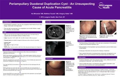
Case Description/Methods: A 16-year-old previously healthy female presented with one week of epigastric pain and intractable nonbloody, nonbilious emesis. Abdominal ultrasound demonstrated small gallstones, but no biliary ductal dilatation or gallbladder wall thickening. MRCP demonstrated severe necrotizing pancreatitis, a thick-walled, septated collection anterior to the pancreas, and a duplication cyst measuring 5.2 x 2.3 x 3.8 cm in the second portion of the duodenum (Figure 1). Also, a large pseudocyst was found on an endosonographic exam of the peritoneal cavity. This was corroborated on EUS (Figure 2). EUS with cystogastrostomy was performed via 15 mm x 15 mm AXIOS stent placement for endoscopic management of the pseudocyst.
Four days post EUS, she was able to tolerate a liquid diet and was discharged with oral antibiotics. During repeat ERCP one month later, the AXIOS stent was removed and the duplication cyst was marsupialized (Figure 3). For safety, a plastic stent was placed in the common bile duct and another was placed in the ventral pancreatic duct. She was sent home with an antibiotic course and we plan to repeat an ERCP with stent removal in eight weeks.
Discussion: There is limited discussion in the literature about pediatric duplication cysts due to their rare occurrence and varied presentations. The likely etiology of pancreatitis in this case was compression of the ampulla of Vater by the duodenal duplication cyst. In recent times, endoscopic intervention has become the mainstay of treatment. In this case, marsupialization of the duplication was performed to prevent further bouts of pancreatitis. However, open surgical intervention is employed when endoscopic management is not possible, such as cyst involvement of the pancreatic head and biliary tract.

Disclosures:
Ria Minawala, MD1, Matthew Fasullo, DO2, Gregory Haber, MD3. P2951 - Periampullary Duodenal Duplication Cyst: An Unsuspecting Cause of Acute Pancreatitis, ACG 2023 Annual Scientific Meeting Abstracts. Vancouver, BC, Canada: American College of Gastroenterology.
1NYU Langone Health, New York, NY; 2NYU Langone Medical Center, New York, NY; 3NYU, New York, NY
Introduction: Gastrointestinal duplication cysts are rare congenital malformations found in the foregut, small bowel, and large bowel. Of these locations, duodenal duplication cysts are exceedingly rare and have a prevalence of less than 1 per 100,000 live births. Although often asymptomatic, complications of duplication cysts most commonly arise in early childhood. Notable manifestations include infection, weight loss, GI bleeding, and pancreatitis. We report a case of a pediatric patient with acute pancreatitis caused by a periampullary duplication cyst and complicated by a pancreatic pseudocyst that were both managed endoscopically.
Case Description/Methods: A 16-year-old previously healthy female presented with one week of epigastric pain and intractable nonbloody, nonbilious emesis. Abdominal ultrasound demonstrated small gallstones, but no biliary ductal dilatation or gallbladder wall thickening. MRCP demonstrated severe necrotizing pancreatitis, a thick-walled, septated collection anterior to the pancreas, and a duplication cyst measuring 5.2 x 2.3 x 3.8 cm in the second portion of the duodenum (Figure 1). Also, a large pseudocyst was found on an endosonographic exam of the peritoneal cavity. This was corroborated on EUS (Figure 2). EUS with cystogastrostomy was performed via 15 mm x 15 mm AXIOS stent placement for endoscopic management of the pseudocyst.
Four days post EUS, she was able to tolerate a liquid diet and was discharged with oral antibiotics. During repeat ERCP one month later, the AXIOS stent was removed and the duplication cyst was marsupialized (Figure 3). For safety, a plastic stent was placed in the common bile duct and another was placed in the ventral pancreatic duct. She was sent home with an antibiotic course and we plan to repeat an ERCP with stent removal in eight weeks.
Discussion: There is limited discussion in the literature about pediatric duplication cysts due to their rare occurrence and varied presentations. The likely etiology of pancreatitis in this case was compression of the ampulla of Vater by the duodenal duplication cyst. In recent times, endoscopic intervention has become the mainstay of treatment. In this case, marsupialization of the duplication was performed to prevent further bouts of pancreatitis. However, open surgical intervention is employed when endoscopic management is not possible, such as cyst involvement of the pancreatic head and biliary tract.

Figure: Figure 1: Duodenal duplication cyst measuring 5.2 x 2.3 x 3.8 cm on MRCP.
Figure 2: Pancreatic pseudocyst seen on EUS.
Figure 3: Duplication cyst visualized at the second portion of the duodenum during ERCP.
Figure 2: Pancreatic pseudocyst seen on EUS.
Figure 3: Duplication cyst visualized at the second portion of the duodenum during ERCP.
Disclosures:
Ria Minawala indicated no relevant financial relationships.
Matthew Fasullo indicated no relevant financial relationships.
Gregory Haber: BSC – Consultant. Digma – Consultant. Erbe – Consultant. Medtronic – Consultant. Microtech – Consultant. Olympus – Consultant. Ovesco – Consultant.
Ria Minawala, MD1, Matthew Fasullo, DO2, Gregory Haber, MD3. P2951 - Periampullary Duodenal Duplication Cyst: An Unsuspecting Cause of Acute Pancreatitis, ACG 2023 Annual Scientific Meeting Abstracts. Vancouver, BC, Canada: American College of Gastroenterology.
