Tuesday Poster Session
Category: Liver
P3958 - A Rare Case of an Intrahepatic Portosystemic Fistula
Tuesday, October 24, 2023
10:30 AM - 4:00 PM PT
Location: Exhibit Hall

Has Audio

Marisa Pope, DO
Jefferson Health
Stratford, NJ
Presenting Author(s)
Marisa Pope, DO1, Saurabh Dharmadhikari, DO1, Joshua Soliman, DO2, C Jonathan Foster, DO3
1Jefferson Health, Stratford, NJ; 2Thomas Jefferson University, Voorhees, NJ; 3Jefferson Health, Cherry Hill, NJ
Introduction: A venous fistula occurs when there is communication between two neighboring veins. Some etiologies of venous fistulas include, iatrogenic, ruptured aneurysm, or congenital. Patients with liver cirrhosis are more prone to developing portosystemic fistulas, which contribute to the development of portal hypertension. A portosystemic fistula is a rare entity. We present a unique case of portohepatic venous fistula in a patient with portal hypertension.
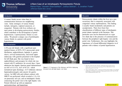
Case Description/Methods: A woman in her 50s with a past medical history of HTN, SVT, paroxysmal atrial fibrillation, nephrolithiasis, hepatitis A, gastric sleeve, cervical laminectomy and recent upper endoscopy with gastritis presented to the hospital for left flank pain. She was found to have nephrolithiasis and urosepsis for which she was treated with IV antibiotics for several days. Hepatic function panel during admission showed AST 112 U/L, ALT 92U/L on admission, which trended down to normal after discharge. CT abdomen showed hepatosplenomegaly and concern for gastric varices. MRI w/wo contrast with MRCP was performed which revealed a 1.8 x 0.6cm pancreatic tail cyst, and an intrahepatic fistula connecting the portal vein to the right hepatic vein. Patient followed up with an outpatient GI physician to establish care.
Discussion: Portosystemic shunts within the liver are a rare occurrence that is commonly associated with congenital venous malformations. This finding is usually found incidentally and patients are asymptomatic. There have been four classifications of different types of intrahepatic portal shunts reported in the literature. This particular case can be characterized as a type two shunt due to the presence of communication between the peripheral right hepatic vein and the right portal vein.Our case is unique as a fistula formed between the right hepatic vein and portal vein. It is important to include in the differential diagnosis of patients with evidence of portal hypertension.

Disclosures:
Marisa Pope, DO1, Saurabh Dharmadhikari, DO1, Joshua Soliman, DO2, C Jonathan Foster, DO3. P3958 - A Rare Case of an Intrahepatic Portosystemic Fistula, ACG 2023 Annual Scientific Meeting Abstracts. Vancouver, BC, Canada: American College of Gastroenterology.
1Jefferson Health, Stratford, NJ; 2Thomas Jefferson University, Voorhees, NJ; 3Jefferson Health, Cherry Hill, NJ
Introduction: A venous fistula occurs when there is communication between two neighboring veins. Some etiologies of venous fistulas include, iatrogenic, ruptured aneurysm, or congenital. Patients with liver cirrhosis are more prone to developing portosystemic fistulas, which contribute to the development of portal hypertension. A portosystemic fistula is a rare entity. We present a unique case of portohepatic venous fistula in a patient with portal hypertension.
Case Description/Methods: A woman in her 50s with a past medical history of HTN, SVT, paroxysmal atrial fibrillation, nephrolithiasis, hepatitis A, gastric sleeve, cervical laminectomy and recent upper endoscopy with gastritis presented to the hospital for left flank pain. She was found to have nephrolithiasis and urosepsis for which she was treated with IV antibiotics for several days. Hepatic function panel during admission showed AST 112 U/L, ALT 92U/L on admission, which trended down to normal after discharge. CT abdomen showed hepatosplenomegaly and concern for gastric varices. MRI w/wo contrast with MRCP was performed which revealed a 1.8 x 0.6cm pancreatic tail cyst, and an intrahepatic fistula connecting the portal vein to the right hepatic vein. Patient followed up with an outpatient GI physician to establish care.
Discussion: Portosystemic shunts within the liver are a rare occurrence that is commonly associated with congenital venous malformations. This finding is usually found incidentally and patients are asymptomatic. There have been four classifications of different types of intrahepatic portal shunts reported in the literature. This particular case can be characterized as a type two shunt due to the presence of communication between the peripheral right hepatic vein and the right portal vein.Our case is unique as a fistula formed between the right hepatic vein and portal vein. It is important to include in the differential diagnosis of patients with evidence of portal hypertension.

Figure: MRI image displaying the right hepatic vein fistula with the right portal vein.
Disclosures:
Marisa Pope indicated no relevant financial relationships.
Saurabh Dharmadhikari indicated no relevant financial relationships.
Joshua Soliman indicated no relevant financial relationships.
C Jonathan Foster indicated no relevant financial relationships.
Marisa Pope, DO1, Saurabh Dharmadhikari, DO1, Joshua Soliman, DO2, C Jonathan Foster, DO3. P3958 - A Rare Case of an Intrahepatic Portosystemic Fistula, ACG 2023 Annual Scientific Meeting Abstracts. Vancouver, BC, Canada: American College of Gastroenterology.
