Tuesday Poster Session
Category: Biliary/Pancreas
P2972 - Multiple Scans, No Answers: Failure of CTs to Detect Symptomatic Pancreatic Cancer
Tuesday, October 24, 2023
10:30 AM - 4:00 PM PT
Location: Exhibit Hall

Has Audio
- KG
Kevin Groudan, MD
University of Massachusetts Chan Medical School-Baystate
Springfield, MA
Presenting Author(s)
Kevin Groudan, MD, Yesenia Greeff, MD
University of Massachusetts Chan Medical School-Baystate, Springfield, MA
Introduction: Pancreatic cancer is the fourth leading cause of cancer death in the United States. Pancreatic protocol computed tomography (CT) with intravenous (IV) contrast is considered standard for the diagnosis of pancreatic cancer. We report a patient with malignant gastric outlet obstruction who had multiple CTs that failed to diagnose pancreatic cancer leading to a delay in diagnosis.
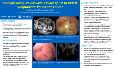
Case Description/Methods: A 67-year-old man presented to gastroenterology clinic with 3 months of epigastric pain associated with a 15-pound weight loss and acid reflux symptoms. He previously went to the emergency room and had an abdominal CT with IV contrast that reported subtle acute pancreatitis and a mildly prominent common bile duct. Complete blood count and complete metabolic panel were normal and lipase was 85 units/L. He was treated with pantoprazole. EGD showed a mild distal esophageal stricture, severe esophagitis, and food in the stomach and duodenal sweep prohibiting examination of the second part of the duodenum. Pantoprazole was increased to twice daily and a repeat EGD was scheduled in 8 weeks after 2 days of a liquid diet. Repeat EGD showed worsened esophagitis, a massively distended stomach, and again food obstructing the duodenal sweep. Abdominal CT with IV contrast was repeated and showed mild intra and extra-hepatic biliary duct dilation and a severely distended stomach. The pancreas was reported as normal.
He was hospitalized for a repeat EGD after nasogastric tube decompression. Repeat EGD showed extrinsic compression in the second part of the duodenum. Biopsies were consistent with a reactive process. Abdominal magnetic resonance imaging showed an ill-defined pancreatic head mass measuring 3.0 x 2.0 cm. Endoscopic ultrasound confirmed presence of the pancreatic head lesion. Fine needle biopsies were consistent with invasive adenocarcinoma of the pancreas. He was referred to surgical oncology and underwent a gastric and biliary bypass surgery and started on chemotherapy.
Discussion: Sensitivity of CT for detection of pancreatic cancer is reported to be between 68 to 77% for tumors less than 2 cm and as high as 98% for tumors greater than 2 cm. Kang et. al found that most missed cases of pancreatic cancer were either less than 2 cm, isoattenuating or non-contour deforming on CT and most misinterpreted cases were reported as pancreatitis. Clinicians should be aware that pancreatic cancer can be missed or misinterpreted on CT.
Disclosures:
Kevin Groudan, MD, Yesenia Greeff, MD. P2972 - Multiple Scans, No Answers: Failure of CTs to Detect Symptomatic Pancreatic Cancer, ACG 2023 Annual Scientific Meeting Abstracts. Vancouver, BC, Canada: American College of Gastroenterology.
University of Massachusetts Chan Medical School-Baystate, Springfield, MA
Introduction: Pancreatic cancer is the fourth leading cause of cancer death in the United States. Pancreatic protocol computed tomography (CT) with intravenous (IV) contrast is considered standard for the diagnosis of pancreatic cancer. We report a patient with malignant gastric outlet obstruction who had multiple CTs that failed to diagnose pancreatic cancer leading to a delay in diagnosis.
Case Description/Methods: A 67-year-old man presented to gastroenterology clinic with 3 months of epigastric pain associated with a 15-pound weight loss and acid reflux symptoms. He previously went to the emergency room and had an abdominal CT with IV contrast that reported subtle acute pancreatitis and a mildly prominent common bile duct. Complete blood count and complete metabolic panel were normal and lipase was 85 units/L. He was treated with pantoprazole. EGD showed a mild distal esophageal stricture, severe esophagitis, and food in the stomach and duodenal sweep prohibiting examination of the second part of the duodenum. Pantoprazole was increased to twice daily and a repeat EGD was scheduled in 8 weeks after 2 days of a liquid diet. Repeat EGD showed worsened esophagitis, a massively distended stomach, and again food obstructing the duodenal sweep. Abdominal CT with IV contrast was repeated and showed mild intra and extra-hepatic biliary duct dilation and a severely distended stomach. The pancreas was reported as normal.
He was hospitalized for a repeat EGD after nasogastric tube decompression. Repeat EGD showed extrinsic compression in the second part of the duodenum. Biopsies were consistent with a reactive process. Abdominal magnetic resonance imaging showed an ill-defined pancreatic head mass measuring 3.0 x 2.0 cm. Endoscopic ultrasound confirmed presence of the pancreatic head lesion. Fine needle biopsies were consistent with invasive adenocarcinoma of the pancreas. He was referred to surgical oncology and underwent a gastric and biliary bypass surgery and started on chemotherapy.
Discussion: Sensitivity of CT for detection of pancreatic cancer is reported to be between 68 to 77% for tumors less than 2 cm and as high as 98% for tumors greater than 2 cm. Kang et. al found that most missed cases of pancreatic cancer were either less than 2 cm, isoattenuating or non-contour deforming on CT and most misinterpreted cases were reported as pancreatitis. Clinicians should be aware that pancreatic cancer can be missed or misinterpreted on CT.
Disclosures:
Kevin Groudan indicated no relevant financial relationships.
Yesenia Greeff indicated no relevant financial relationships.
Kevin Groudan, MD, Yesenia Greeff, MD. P2972 - Multiple Scans, No Answers: Failure of CTs to Detect Symptomatic Pancreatic Cancer, ACG 2023 Annual Scientific Meeting Abstracts. Vancouver, BC, Canada: American College of Gastroenterology.
