Tuesday Poster Session
Category: Biliary/Pancreas
P2976 - DIY Meets WOPN: Spontaneous Erosion of a Peripancreatic Fluid Collection Into the Stomach
Tuesday, October 24, 2023
10:30 AM - 4:00 PM PT
Location: Exhibit Hall

Has Audio
- MP
Madison Peregoy, MD
National Capital Consortium
Bethesda, MD
Presenting Author(s)
Madison Peregoy, MD1, Andrew Mertz, MD2, Patrick Young, MD, FACG2
1National Capital Consortium, Bethesda, MD; 2Walter Reed National Military Medical Center, Bethesda, MD
Introduction: Pancreatic fluid collections are a common complication of both interstitial and necrotizing pancreatitis. Drainage is indicated if there is a concern for superinfection or symptoms secondary to size or location. The proximity of the pancreas to the stomach and duodenum makes stenting the top therapeutic choice for drainage in many cases. Uncommonly, fluid collections can enlarge and erode into nearby structures. Herein, we describe a case of spontaneous infected pancreatic fluid collection decompression via gastric fistula.
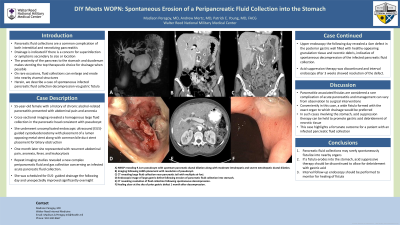
Case Description/Methods: A 55-year-old female with a history of chronic alcohol-related pancreatitis presented with abdominal pain and anorexia. Cross-sectional imaging revealed a homogenous large fluid collection located in the pancreatic head consistent with pseudocyst, which was followed with repeat imaging studies. She subsequently underwent uncomplicated endoscopic ultrasound (EUS) guided cystoduodenostomy with placement of a lumen opposing metal stent along with common bile duct stent placement for biliary obstruction. One month later she represented with recurrent abdominal pain, anorexia, fever, and leukocytosis. Repeat imaging studies revealed a new complex peripancreatic fluid and gas collection concerning an infected acute pancreatic fluid collection. She was scheduled for EUS- guided drainage the following day and interestingly improved symptomatically overnight just prior to the procedure. Upper endoscopy the following day revealed a 4cm defect in the posterior gastric wall filled with healthy-appearing granulation tissue and necrotic debris, indicative of spontaneous decompression of the infected pancreatic fluid collection. Acid suppression therapy was discontinued and interval endoscopy after 3 weeks showed resolution of the defect.
Discussion: Pancreatitis-associated fistulas are considered a rare complication of acute pancreatitis and management can vary from observation to surgical interventions. Conveniently in this case, a wide fistula formed with the exact organ to which drainage would be preferred. In such cases involving the stomach, acid suppression therapy can be held to promote gastric acid debridement of necrotic tissue, allowing the fistula to remain patent for adequate drainage. This case highlights a fortunate outcome for a patient with an infected pancreatic fluid collection, however it also reminds us to respect such fluid collections as they can hastily evolve.

Disclosures:
Madison Peregoy, MD1, Andrew Mertz, MD2, Patrick Young, MD, FACG2. P2976 - DIY Meets WOPN: Spontaneous Erosion of a Peripancreatic Fluid Collection Into the Stomach, ACG 2023 Annual Scientific Meeting Abstracts. Vancouver, BC, Canada: American College of Gastroenterology.
1National Capital Consortium, Bethesda, MD; 2Walter Reed National Military Medical Center, Bethesda, MD
Introduction: Pancreatic fluid collections are a common complication of both interstitial and necrotizing pancreatitis. Drainage is indicated if there is a concern for superinfection or symptoms secondary to size or location. The proximity of the pancreas to the stomach and duodenum makes stenting the top therapeutic choice for drainage in many cases. Uncommonly, fluid collections can enlarge and erode into nearby structures. Herein, we describe a case of spontaneous infected pancreatic fluid collection decompression via gastric fistula.
Case Description/Methods: A 55-year-old female with a history of chronic alcohol-related pancreatitis presented with abdominal pain and anorexia. Cross-sectional imaging revealed a homogenous large fluid collection located in the pancreatic head consistent with pseudocyst, which was followed with repeat imaging studies. She subsequently underwent uncomplicated endoscopic ultrasound (EUS) guided cystoduodenostomy with placement of a lumen opposing metal stent along with common bile duct stent placement for biliary obstruction. One month later she represented with recurrent abdominal pain, anorexia, fever, and leukocytosis. Repeat imaging studies revealed a new complex peripancreatic fluid and gas collection concerning an infected acute pancreatic fluid collection. She was scheduled for EUS- guided drainage the following day and interestingly improved symptomatically overnight just prior to the procedure. Upper endoscopy the following day revealed a 4cm defect in the posterior gastric wall filled with healthy-appearing granulation tissue and necrotic debris, indicative of spontaneous decompression of the infected pancreatic fluid collection. Acid suppression therapy was discontinued and interval endoscopy after 3 weeks showed resolution of the defect.
Discussion: Pancreatitis-associated fistulas are considered a rare complication of acute pancreatitis and management can vary from observation to surgical interventions. Conveniently in this case, a wide fistula formed with the exact organ to which drainage would be preferred. In such cases involving the stomach, acid suppression therapy can be held to promote gastric acid debridement of necrotic tissue, allowing the fistula to remain patent for adequate drainage. This case highlights a fortunate outcome for a patient with an infected pancreatic fluid collection, however it also reminds us to respect such fluid collections as they can hastily evolve.

Figure: A. MRCP revealing 9.1cm pseudocyst with upstream pancreatic ductal dilation along with moderate intrahepatic and severe extrahepatic ductal dilation.
B. Imaging following LAMS placement with resolution of pseudocyst.
C. CT revealing large fluid collection near pancreatic tail with multiple air foci.
D. Endoscopic image of large gastric defect following erosion of pancreatic fluid collection into stomach.
E. CT revealing resolution of fluid collection following spontaneous decompression.
F. Healing ulcer at the site of prior gastric defect 1 month after decompression.
Disclosures:
Madison Peregoy indicated no relevant financial relationships.
Andrew Mertz indicated no relevant financial relationships.
Patrick Young indicated no relevant financial relationships.
Madison Peregoy, MD1, Andrew Mertz, MD2, Patrick Young, MD, FACG2. P2976 - DIY Meets WOPN: Spontaneous Erosion of a Peripancreatic Fluid Collection Into the Stomach, ACG 2023 Annual Scientific Meeting Abstracts. Vancouver, BC, Canada: American College of Gastroenterology.

