Tuesday Poster Session
Category: Biliary/Pancreas
P2984 - Intrabiliary Metastases of Colorectal Cancer: An Under Recognized Entity
Tuesday, October 24, 2023
10:30 AM - 4:00 PM PT
Location: Exhibit Hall

Has Audio
- SY
Sujani Yadlapati, MD
Cooper Medical School of Rowan University/Cooper University Hospital
Camden, New Jersey
Presenting Author(s)
Sujani Yadlapati, MD1, Michael Garrett House, MD2, Nikolay Popnikolov, MD, PHD2, Evan Fogel, MD3
1Cooper Medical School of Rowan University/Cooper University Hospital, Camden, NJ; 2Indiana University Health, Indianapolis, IN; 3Indiana University School of Medicine, Indianapolis, IN
Introduction: Colorectal cancer is prone to distant metastases. Biliary obstruction, when present, is typically due to hepatic metastases or porta hepatis lymph nodes causing extrinsic biliary compression. In rare cases, however, metastatic colorectal cancer can infiltrate the biliary epithelium and spread within the bile ducts, resulting in intrabiliary metastasis. Here, we present two cases of intrabiliary metastases in patients with a prior history of colorectal cancer.
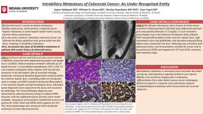
Case Description/Methods: 1. An 84-year-old man with history of colon cancer, resected in 08/2019, presented with abdominal discomfort and weight loss in 12/2022. Initial evaluation revealed a bilirubin of 2.8 mg/dl (normal < 1) and alkaline phosphatase 218 (< 125). CT scan showed intrahepatic biliary dilation with low-density structures in the left hepatic lobe of uncertain etiology. Endoscopic ultrasound identified hyperechoic material within the common hepatic duct, resembling either an intraductal mass or sludge, and ERCP revealed intraductal filling defects in the common hepatic and left intrahepatic ducts. Abundant tumor fragments were swept from the ducts and recovered for pathology. The initial pathologic diagnosis was interpreted as adenocarcinoma arising in a biliary IPMN. However, with the additional history of colon cancer made available, subsequent immunostains revealed the tumor to be positive for CK20, CDX2 and SATB2 while negative for CK7. This immunophenotype was consistent with intraductal metastasis of colon adenocarcinoma.
2. This 68-year-old woman with a history of rectal cancer resected in 2020 presented in 05/2023 with abdominal and back pain and jaundice (bilirubin 17.5 mg/dl). CT scan revealed a porta hepatic mass with bilateral intrahepatic biliary dilation. ERCP revealed filling defects of the common hepatic duct, right intrahepatic ducts and gallbladder, and abundant polypoid filling defects were swept from the duct. Pathology was consistent with adenocarcinoma, and Immunostains revealed the tumor cells to be positive for SATB2 and negative for CK7 and CK20, consistent with intestinal origin.
Discussion: Intrabiliary metastasis in colorectal cancer has been described previously, with experience typically limited to case reports. While cross-sectional imaging aids in evaluation, differentiation from other biliary tumors can be challenging. Clinical suspicion is required, and confirmation requires histopathological evaluation with immunostains for accurate diagnosis.

Disclosures:
Sujani Yadlapati, MD1, Michael Garrett House, MD2, Nikolay Popnikolov, MD, PHD2, Evan Fogel, MD3. P2984 - Intrabiliary Metastases of Colorectal Cancer: An Under Recognized Entity, ACG 2023 Annual Scientific Meeting Abstracts. Vancouver, BC, Canada: American College of Gastroenterology.
1Cooper Medical School of Rowan University/Cooper University Hospital, Camden, NJ; 2Indiana University Health, Indianapolis, IN; 3Indiana University School of Medicine, Indianapolis, IN
Introduction: Colorectal cancer is prone to distant metastases. Biliary obstruction, when present, is typically due to hepatic metastases or porta hepatis lymph nodes causing extrinsic biliary compression. In rare cases, however, metastatic colorectal cancer can infiltrate the biliary epithelium and spread within the bile ducts, resulting in intrabiliary metastasis. Here, we present two cases of intrabiliary metastases in patients with a prior history of colorectal cancer.
Case Description/Methods: 1. An 84-year-old man with history of colon cancer, resected in 08/2019, presented with abdominal discomfort and weight loss in 12/2022. Initial evaluation revealed a bilirubin of 2.8 mg/dl (normal < 1) and alkaline phosphatase 218 (< 125). CT scan showed intrahepatic biliary dilation with low-density structures in the left hepatic lobe of uncertain etiology. Endoscopic ultrasound identified hyperechoic material within the common hepatic duct, resembling either an intraductal mass or sludge, and ERCP revealed intraductal filling defects in the common hepatic and left intrahepatic ducts. Abundant tumor fragments were swept from the ducts and recovered for pathology. The initial pathologic diagnosis was interpreted as adenocarcinoma arising in a biliary IPMN. However, with the additional history of colon cancer made available, subsequent immunostains revealed the tumor to be positive for CK20, CDX2 and SATB2 while negative for CK7. This immunophenotype was consistent with intraductal metastasis of colon adenocarcinoma.
2. This 68-year-old woman with a history of rectal cancer resected in 2020 presented in 05/2023 with abdominal and back pain and jaundice (bilirubin 17.5 mg/dl). CT scan revealed a porta hepatic mass with bilateral intrahepatic biliary dilation. ERCP revealed filling defects of the common hepatic duct, right intrahepatic ducts and gallbladder, and abundant polypoid filling defects were swept from the duct. Pathology was consistent with adenocarcinoma, and Immunostains revealed the tumor cells to be positive for SATB2 and negative for CK7 and CK20, consistent with intestinal origin.
Discussion: Intrabiliary metastasis in colorectal cancer has been described previously, with experience typically limited to case reports. While cross-sectional imaging aids in evaluation, differentiation from other biliary tumors can be challenging. Clinical suspicion is required, and confirmation requires histopathological evaluation with immunostains for accurate diagnosis.

Figure: Fluoroscopy Images 1 and 2: Intraductal filling defects in the common bile duct .
Pathology : A) low grade adenocarcinoma with cribriform and papillary patterns. B) Tumor cells negative for CK7. C)Tumor cells positive for CK20. D) Tumor cells positive for SATB2. This immunophenotype is consistent with a colonic origin.
Pathology : A) low grade adenocarcinoma with cribriform and papillary patterns. B) Tumor cells negative for CK7. C)Tumor cells positive for CK20. D) Tumor cells positive for SATB2. This immunophenotype is consistent with a colonic origin.
Disclosures:
Sujani Yadlapati indicated no relevant financial relationships.
Michael Garrett House indicated no relevant financial relationships.
Nikolay Popnikolov indicated no relevant financial relationships.
Evan Fogel indicated no relevant financial relationships.
Sujani Yadlapati, MD1, Michael Garrett House, MD2, Nikolay Popnikolov, MD, PHD2, Evan Fogel, MD3. P2984 - Intrabiliary Metastases of Colorectal Cancer: An Under Recognized Entity, ACG 2023 Annual Scientific Meeting Abstracts. Vancouver, BC, Canada: American College of Gastroenterology.
