Tuesday Poster Session
Category: Liver
P3992 - A Rare Case of Saddle Pulmonary Embolism Secondary to Hemorrhagic Hepatic Cyst
Tuesday, October 24, 2023
10:30 AM - 4:00 PM PT
Location: Exhibit Hall

Has Audio

Shazia Rashid, MD
LSU Health Sciences Center
Shreveport, LA
Presenting Author(s)
Shazia Rashid, MD1, Daniyal Raza, MD1, Omar Khan, MD1, Sudha Pandit, MD1, Natalie Bzowej, MD, PhD2
1LSU Health Sciences Center, Shreveport, LA; 2Ochsner, New Orleans, LA
Introduction: Nonparasitic hepatic cysts are considered rare, with an estimated global prevalence of only 5%. These cysts are characterized by fluid-filled cavities in the liver, appear asymptomatic and generally do not necessitate treatment unless cause rupture. This is a case of an elderly female patient with a saddle pulmonary embolism (PE) secondary to large hepatic cyst.
Case Description/Methods: A 76-year-old female with a history of hepatic cysts presented with severe right upper quadrant abdominal pain and shortness of breath lasting for two days. She had been diagnosed with liver cysts at the age of 40 and had undergone ultrasound surveillance every five years. Her last assessment few months prior to presentation revealed an enlarging asymptomatic cyst from 8.5cm to 17cm. Further investigations at that time ruled out cirrhosis, fatty liver, and hepatitis; Ca-125 level was normal. On examination, abdominal fullness was noted. A computed tomography angiogram (CTA) of the chest showed non obstructive pulmonary embolism and 21cm hepatic cyst. Anticoagulation was started with development of an acute blood loss anemia requiring transfusions. Subsequent CT abdomen revealed hyperdensity in the cyst within venous phase likely venous bleeding. Multiple angiograms revealed no active arterial extravasation and thus no embolization was performed. Given patient’s BMI, she was deemed a high surgical candidate. It was postulated that the low venous flow and inferior vena cava (IVC) compression caused by the cysts may have contributed to the development of the pulmonary embolism. Subsequently, patient underwent IR drainage of hepatic cyst with resultant 200cc of dark blood aspirated. She continues to follow up with IR and hepatology outpatient.
Discussion: Simple hepatic cysts are usually asymptomatic and incidentally diagnosed. They can vary in size, ranging from a few millimeters to enormous proportions, with the right hepatic lobe being the most common location. Laboratory test results are often normal. Sonography, Computed Tomography, or magnetic resonance imaging are used to detect and survey these cysts. This case is one of the few reported instances where an abdominal cyst compromised the inferior vena cava (IVC), leading to pulmonary embolism. While most hepatic cysts do not cause symptoms, complications such as hemorrhage within the abdominal cavity and infections can occur. Treatment options for symptomatic patients may involve percutaneous drainage, open or laparoscopic deroofing, or hepatic resection.

Disclosures:
Shazia Rashid, MD1, Daniyal Raza, MD1, Omar Khan, MD1, Sudha Pandit, MD1, Natalie Bzowej, MD, PhD2. P3992 - A Rare Case of Saddle Pulmonary Embolism Secondary to Hemorrhagic Hepatic Cyst, ACG 2023 Annual Scientific Meeting Abstracts. Vancouver, BC, Canada: American College of Gastroenterology.
1LSU Health Sciences Center, Shreveport, LA; 2Ochsner, New Orleans, LA
Introduction: Nonparasitic hepatic cysts are considered rare, with an estimated global prevalence of only 5%. These cysts are characterized by fluid-filled cavities in the liver, appear asymptomatic and generally do not necessitate treatment unless cause rupture. This is a case of an elderly female patient with a saddle pulmonary embolism (PE) secondary to large hepatic cyst.
Case Description/Methods: A 76-year-old female with a history of hepatic cysts presented with severe right upper quadrant abdominal pain and shortness of breath lasting for two days. She had been diagnosed with liver cysts at the age of 40 and had undergone ultrasound surveillance every five years. Her last assessment few months prior to presentation revealed an enlarging asymptomatic cyst from 8.5cm to 17cm. Further investigations at that time ruled out cirrhosis, fatty liver, and hepatitis; Ca-125 level was normal. On examination, abdominal fullness was noted. A computed tomography angiogram (CTA) of the chest showed non obstructive pulmonary embolism and 21cm hepatic cyst. Anticoagulation was started with development of an acute blood loss anemia requiring transfusions. Subsequent CT abdomen revealed hyperdensity in the cyst within venous phase likely venous bleeding. Multiple angiograms revealed no active arterial extravasation and thus no embolization was performed. Given patient’s BMI, she was deemed a high surgical candidate. It was postulated that the low venous flow and inferior vena cava (IVC) compression caused by the cysts may have contributed to the development of the pulmonary embolism. Subsequently, patient underwent IR drainage of hepatic cyst with resultant 200cc of dark blood aspirated. She continues to follow up with IR and hepatology outpatient.
Discussion: Simple hepatic cysts are usually asymptomatic and incidentally diagnosed. They can vary in size, ranging from a few millimeters to enormous proportions, with the right hepatic lobe being the most common location. Laboratory test results are often normal. Sonography, Computed Tomography, or magnetic resonance imaging are used to detect and survey these cysts. This case is one of the few reported instances where an abdominal cyst compromised the inferior vena cava (IVC), leading to pulmonary embolism. While most hepatic cysts do not cause symptoms, complications such as hemorrhage within the abdominal cavity and infections can occur. Treatment options for symptomatic patients may involve percutaneous drainage, open or laparoscopic deroofing, or hepatic resection.

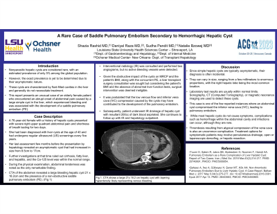
Figure: A Contrast-enhanced Computed Tomographic image of a large hepatic cyst with layering hyperintense material suggesting bleeding within the cyst.
Disclosures:
Shazia Rashid indicated no relevant financial relationships.
Daniyal Raza indicated no relevant financial relationships.
Omar Khan indicated no relevant financial relationships.
Sudha Pandit indicated no relevant financial relationships.
Natalie Bzowej indicated no relevant financial relationships.
Shazia Rashid, MD1, Daniyal Raza, MD1, Omar Khan, MD1, Sudha Pandit, MD1, Natalie Bzowej, MD, PhD2. P3992 - A Rare Case of Saddle Pulmonary Embolism Secondary to Hemorrhagic Hepatic Cyst, ACG 2023 Annual Scientific Meeting Abstracts. Vancouver, BC, Canada: American College of Gastroenterology.
