Tuesday Poster Session
Category: Liver
P3993 - Recurrent HCC Masquerading as Nodular GAVE in a Patient With Prior Cryoablation
Tuesday, October 24, 2023
10:30 AM - 4:00 PM PT
Location: Exhibit Hall

Has Audio

Katherine M. Cooper, MD
UMass Chan Medical School
Worcester, MA
Presenting Author(s)
Katherine M.. Cooper, MD1, Peter Sullivan, MD1, Savant Mehta, MBBS, MD, DM2, Deepika Devuni, MD1
1UMass Chan Medical School, Worcester, MA; 2UMass Memorial Healthcare, Worcester, MA
Introduction: Recurrent hepatocellular carcinoma (rHCC) is not uncommon. Extrahepatic recurrence (EHR) is common after liver transplant (LT) while intrahepatic recurrence (IHR) occurs more after ablations (1,2). We here-in present an unusual case of rHCC presenting as a gastric mass.
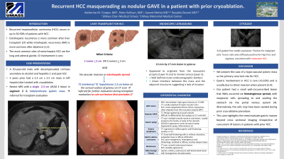
Case Description/Methods: A 63-year-old male with alcohol and hepatitis C associated cirrhosis who presents for endoscopic evaluation of a gastric mass identified during LT evaluation (LTE). Five years prior, he underwent cryoablation for a 2.5 x 2.5 cm biopsy-confirmed HCC in the left hepatic lobe. Surveillance scans since that time were stable until MRI obtained during LTE showed a new 2.4 cm LiRADS-4 lesion and a T1 isointense/T2 hypointense 1.3 cm lesion of unclear etiology arising from the serosal surface of pylorus. Review of prior CT scans showed correlation with a small lesion attributed to be known nodular GAVE, though the differential for the mass also included other benign nodular issue, a reactive lymph node, gastric polyp, adenocarcinoma, and GIST tumor. Endoscopic ultrasound (EUS) was chosen to evaluate the mass prior to pursuing loco-regional therapy. EUS visualized a well circumscribed, intramural (subepithelial) lesion in the prepyloric region of the stomach that appeared to originate from between the muscularis propria (Layer 4) and serosa (Layer 5). There were no vascular structures in the region of the mass. Sonographic appearance was suggestive of a GIST tumor. However, tissue obtained via fine needle aspiration demonstrated tumor cells that were diffusely positive for Hep-Par1 and arginase, consistent with rHCC. In retrospect, the evolution of the mass on prior scans the pyloric lesion was primary EH recurrence site. Due to the advanced nature of the disease, he was removed from LT consideration and referred to oncology. Unfortunately, he did not tolerate systemic therapy and transitioned to hospice.
Discussion: In this case, a hypo-vascular pyloric mass previously attributed to nodular GAVE was ultimately diagnosed as rHCC in a patient with 4 years of stable scans. Gastric involvement in HCC is rare and often involves direct invasion when present (2,3). Our patient had a small well-circumscribed lesion that likely occurred via hematogenous spread, which may seed the stomach via the portal venous system. This case highlights the need evaluate gastric masses including those with potential other causes, like GAVE, beyond cross sectional imaging irrespective of concurrent IH lesions in patients with prior HCC.

Disclosures:
Katherine M.. Cooper, MD1, Peter Sullivan, MD1, Savant Mehta, MBBS, MD, DM2, Deepika Devuni, MD1. P3993 - Recurrent HCC Masquerading as Nodular GAVE in a Patient With Prior Cryoablation, ACG 2023 Annual Scientific Meeting Abstracts. Vancouver, BC, Canada: American College of Gastroenterology.
1UMass Chan Medical School, Worcester, MA; 2UMass Memorial Healthcare, Worcester, MA
Introduction: Recurrent hepatocellular carcinoma (rHCC) is not uncommon. Extrahepatic recurrence (EHR) is common after liver transplant (LT) while intrahepatic recurrence (IHR) occurs more after ablations (1,2). We here-in present an unusual case of rHCC presenting as a gastric mass.
Case Description/Methods: A 63-year-old male with alcohol and hepatitis C associated cirrhosis who presents for endoscopic evaluation of a gastric mass identified during LT evaluation (LTE). Five years prior, he underwent cryoablation for a 2.5 x 2.5 cm biopsy-confirmed HCC in the left hepatic lobe. Surveillance scans since that time were stable until MRI obtained during LTE showed a new 2.4 cm LiRADS-4 lesion and a T1 isointense/T2 hypointense 1.3 cm lesion of unclear etiology arising from the serosal surface of pylorus. Review of prior CT scans showed correlation with a small lesion attributed to be known nodular GAVE, though the differential for the mass also included other benign nodular issue, a reactive lymph node, gastric polyp, adenocarcinoma, and GIST tumor. Endoscopic ultrasound (EUS) was chosen to evaluate the mass prior to pursuing loco-regional therapy. EUS visualized a well circumscribed, intramural (subepithelial) lesion in the prepyloric region of the stomach that appeared to originate from between the muscularis propria (Layer 4) and serosa (Layer 5). There were no vascular structures in the region of the mass. Sonographic appearance was suggestive of a GIST tumor. However, tissue obtained via fine needle aspiration demonstrated tumor cells that were diffusely positive for Hep-Par1 and arginase, consistent with rHCC. In retrospect, the evolution of the mass on prior scans the pyloric lesion was primary EH recurrence site. Due to the advanced nature of the disease, he was removed from LT consideration and referred to oncology. Unfortunately, he did not tolerate systemic therapy and transitioned to hospice.
Discussion: In this case, a hypo-vascular pyloric mass previously attributed to nodular GAVE was ultimately diagnosed as rHCC in a patient with 4 years of stable scans. Gastric involvement in HCC is rare and often involves direct invasion when present (2,3). Our patient had a small well-circumscribed lesion that likely occurred via hematogenous spread, which may seed the stomach via the portal venous system. This case highlights the need evaluate gastric masses including those with potential other causes, like GAVE, beyond cross sectional imaging irrespective of concurrent IH lesions in patients with prior HCC.

Figure: Radiographic and endoscopic evaluation of gastric mass
Disclosures:
Katherine Cooper indicated no relevant financial relationships.
Peter Sullivan indicated no relevant financial relationships.
Savant Mehta indicated no relevant financial relationships.
Deepika Devuni: NIAAA (AA017986) – Grant/Research Support. Sequana Medical – Grant/Research Support.
Katherine M.. Cooper, MD1, Peter Sullivan, MD1, Savant Mehta, MBBS, MD, DM2, Deepika Devuni, MD1. P3993 - Recurrent HCC Masquerading as Nodular GAVE in a Patient With Prior Cryoablation, ACG 2023 Annual Scientific Meeting Abstracts. Vancouver, BC, Canada: American College of Gastroenterology.
