Tuesday Poster Session
Category: Colon
P3059 - Hymenolepis Nana Diagnosed on Colonoscopy in a Renal Transplant Patient
Tuesday, October 24, 2023
10:30 AM - 4:00 PM PT
Location: Exhibit Hall

Has Audio
- KY
Kevin Yang, MD
University at Buffalo
Buffalo, NY
Presenting Author(s)
Kevin Yang, MD, Ali A.. Aijaz, DO, Naren S. Nallapeta, MBBS, Thomas C. Mahl, MD
University at Buffalo, Buffalo, NY
Introduction: Hymenolepsis nana (HN) is the most common cestode infection worldwide and most commonly affects children, people living in institutional settings, and populations lacking proper sanitation. Patients are usually asymptomatic until the degree of parasitic burden becomes severe enough to manifest somatically, particularly as gastrointestinal complaints. This case is unique in that the disease was first identified on colonoscopy in an immunocompromised host.
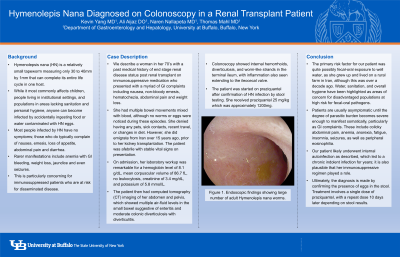
Case Description/Methods: A woman in her 70’s with recent history of renal transplant on immunosuppressive medication presented with nausea, non-bloody emesis, hematochezia, abdominal pain and weight loss. She was in her normal state of health until 2 days prior to hospital admission. She had multiple bowel movements mixed with blood, although no worms or eggs were noticed during these episodes. She denied having any pets, sick contacts, recent travel, or changes in diet. However, she did emigrate from Iran over 15 years ago, prior to her kidney transplantation. She was well appearing on exam without rashes or jaundice. On admission, her laboratory workup was remarkable for a hemoglobin level of 8.1 g/dL without leukocytosis. The patient underwent computed tomography imaging of her abdomen and pelvis, which showed enteritis and colonic diverticulosis. Her upper endoscopy was unremarkable, and colonoscopy showed internal hemorrhoids, diverticulosis, and worm-like strands in the terminal ileum, with inflammation also seen extending to the ileocecal valve (Figure 1). Biopsies were taken around the ileocecal valve and terminal ileum and were remarkable for lymphoid aggregates and fibrinopurulent material consistent with colitis. She subsequently underwent stool ova and parasites testing, which was positive for HN eggs. The patient was started on praziquantel and reported complete resolution of symptoms at her outpatient followup appointment.
Discussion: HN often leads to internal auto-infection, indolently persisting for years without being passed into the stool. The primary risk factor for the patient was fecal-oral exposure to well water, although this was over a decade prior to her renal transplant. Her immunosuppressive regimen may have played a role as well, with the parasitic burden progressively increasing over time through repetitive cycles of tapeworm reproduction. Regardless, this report highlights the utility of colonoscopy to aid in timely diagnosis when initial suspicion for parasitic infection is low.

Disclosures:
Kevin Yang, MD, Ali A.. Aijaz, DO, Naren S. Nallapeta, MBBS, Thomas C. Mahl, MD. P3059 - Hymenolepis Nana Diagnosed on Colonoscopy in a Renal Transplant Patient, ACG 2023 Annual Scientific Meeting Abstracts. Vancouver, BC, Canada: American College of Gastroenterology.
University at Buffalo, Buffalo, NY
Introduction: Hymenolepsis nana (HN) is the most common cestode infection worldwide and most commonly affects children, people living in institutional settings, and populations lacking proper sanitation. Patients are usually asymptomatic until the degree of parasitic burden becomes severe enough to manifest somatically, particularly as gastrointestinal complaints. This case is unique in that the disease was first identified on colonoscopy in an immunocompromised host.
Case Description/Methods: A woman in her 70’s with recent history of renal transplant on immunosuppressive medication presented with nausea, non-bloody emesis, hematochezia, abdominal pain and weight loss. She was in her normal state of health until 2 days prior to hospital admission. She had multiple bowel movements mixed with blood, although no worms or eggs were noticed during these episodes. She denied having any pets, sick contacts, recent travel, or changes in diet. However, she did emigrate from Iran over 15 years ago, prior to her kidney transplantation. She was well appearing on exam without rashes or jaundice. On admission, her laboratory workup was remarkable for a hemoglobin level of 8.1 g/dL without leukocytosis. The patient underwent computed tomography imaging of her abdomen and pelvis, which showed enteritis and colonic diverticulosis. Her upper endoscopy was unremarkable, and colonoscopy showed internal hemorrhoids, diverticulosis, and worm-like strands in the terminal ileum, with inflammation also seen extending to the ileocecal valve (Figure 1). Biopsies were taken around the ileocecal valve and terminal ileum and were remarkable for lymphoid aggregates and fibrinopurulent material consistent with colitis. She subsequently underwent stool ova and parasites testing, which was positive for HN eggs. The patient was started on praziquantel and reported complete resolution of symptoms at her outpatient followup appointment.
Discussion: HN often leads to internal auto-infection, indolently persisting for years without being passed into the stool. The primary risk factor for the patient was fecal-oral exposure to well water, although this was over a decade prior to her renal transplant. Her immunosuppressive regimen may have played a role as well, with the parasitic burden progressively increasing over time through repetitive cycles of tapeworm reproduction. Regardless, this report highlights the utility of colonoscopy to aid in timely diagnosis when initial suspicion for parasitic infection is low.

Figure: Colonoscopy showing large number of adult Hymenolepis nana worms
Disclosures:
Kevin Yang indicated no relevant financial relationships.
Ali Aijaz indicated no relevant financial relationships.
Naren Nallapeta indicated no relevant financial relationships.
Thomas Mahl indicated no relevant financial relationships.
Kevin Yang, MD, Ali A.. Aijaz, DO, Naren S. Nallapeta, MBBS, Thomas C. Mahl, MD. P3059 - Hymenolepis Nana Diagnosed on Colonoscopy in a Renal Transplant Patient, ACG 2023 Annual Scientific Meeting Abstracts. Vancouver, BC, Canada: American College of Gastroenterology.
