Tuesday Poster Session
Category: Small Intestine
P4128 - Sometimes it's Not Cannabis Hyperemesis Syndrome: A Case of Duodenal Adenocarcinoma in a 48-Year-Old Female
Tuesday, October 24, 2023
10:30 AM - 4:00 PM PT
Location: Exhibit Hall

Has Audio

Idrees Suliman, MD
Midwestern University
Mesa, AZ
Presenting Author(s)
Idrees Suliman, MD1, Kristen Farraj, DO2, Howard Chung, MD3, Adnan Malik, MD1, Mukarram Amine, DO1, Zachary M. Hanafi, DO4, Afsheen Moshtaghi, DO5, Sudhakar Reddy, MD1
1Midwestern University, Mesa, AZ; 2Midwestern University, Chandler, AZ; 3Mountain Vista Medical Center, Mesa, AZ; 4Verde Valley Medical Center, Cottonwood, AZ; 5Midwestern University, Cottonwood, AZ
Introduction: Small Bowel Neoplasms are rare tumors and represent only 3% of all gastrointestinal tumors. In the US the incidence of small bowel cancer appears to be increasing. Small bowel adenocarcinoma has a predilection for the duodenum and typically presents in patients aged 50-70. Risk factors associated with Small Bowel Adenocarcinoma include refined sugar, alcohol, smoked foods and chronic inflammation (IBD particularly Crohn's Disease). We present a unique case of duodenal adenocarcinoma that presented in the setting of Ulcerative Colitis, PUD, and H pylori infection.
Case Description/Methods: 48 yo fem presented to the ER with a 1 month history of epigastric pain and nausea in a background significant for daily marijuana use. She was referred to GI and an EGD and Colonoscopy were performed. She was also found to have a normocytic anemia (Hb9.3). Endoscopy showed changes consistent with UC in the rectum (confirmed by biopsy) and a gastric ulcer. Biopsies from the stomach were positive for H Pylori. She was commenced on rectal mesalamine and quadruple therapy for H pylori. In spite of treatment for the above she continued to have progressive symptoms of post prandial pain, nausea, and vomiting for the next two months. She presented to the ER twice with the same symptoms/continued anemia and was discharged home with symptomatic management. CT w/ contrast did not show any acute abnormalities or bowel thickening. On the third presentation to the ER she was admitted for further evaluation. HIDA scan with CCK was unrevealing. Gastric Emptying study showed abnormal prolongation of solid phase gastric emptying. With symptomatic management she was discharged with a plan for repeat EGD. Repeat outpatient EGD showed fungating mass with stigmata of recent bleeding in the third portion of the duodenum(pictured). Biopsies confirmed duodenal adenocarcinoma.
Discussion: When there are signs and symptoms of GOO, a mechanical obstruction must be ruled out with EGD. Duodenal ulcers typically have a low malignant potential especially if an alternative etiology of ulcer is present. This unfortunate individual suffered from nausea and vomiting which could possibly be explained by cannabis and a duodenal ulcer in the setting of H pylori. It seems however her symptoms were in fact related to a duodenal adenocarcinoma. Persistent anemia was the only other indicator of possible malignancy. This case highlights the importance of broad differential diagnosis, avoiding anchoring bias, and high quality endoscopic evaluation.

Disclosures:
Idrees Suliman, MD1, Kristen Farraj, DO2, Howard Chung, MD3, Adnan Malik, MD1, Mukarram Amine, DO1, Zachary M. Hanafi, DO4, Afsheen Moshtaghi, DO5, Sudhakar Reddy, MD1. P4128 - Sometimes it's Not Cannabis Hyperemesis Syndrome: A Case of Duodenal Adenocarcinoma in a 48-Year-Old Female, ACG 2023 Annual Scientific Meeting Abstracts. Vancouver, BC, Canada: American College of Gastroenterology.
1Midwestern University, Mesa, AZ; 2Midwestern University, Chandler, AZ; 3Mountain Vista Medical Center, Mesa, AZ; 4Verde Valley Medical Center, Cottonwood, AZ; 5Midwestern University, Cottonwood, AZ
Introduction: Small Bowel Neoplasms are rare tumors and represent only 3% of all gastrointestinal tumors. In the US the incidence of small bowel cancer appears to be increasing. Small bowel adenocarcinoma has a predilection for the duodenum and typically presents in patients aged 50-70. Risk factors associated with Small Bowel Adenocarcinoma include refined sugar, alcohol, smoked foods and chronic inflammation (IBD particularly Crohn's Disease). We present a unique case of duodenal adenocarcinoma that presented in the setting of Ulcerative Colitis, PUD, and H pylori infection.
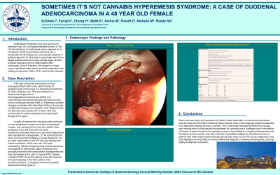
Case Description/Methods: 48 yo fem presented to the ER with a 1 month history of epigastric pain and nausea in a background significant for daily marijuana use. She was referred to GI and an EGD and Colonoscopy were performed. She was also found to have a normocytic anemia (Hb9.3). Endoscopy showed changes consistent with UC in the rectum (confirmed by biopsy) and a gastric ulcer. Biopsies from the stomach were positive for H Pylori. She was commenced on rectal mesalamine and quadruple therapy for H pylori. In spite of treatment for the above she continued to have progressive symptoms of post prandial pain, nausea, and vomiting for the next two months. She presented to the ER twice with the same symptoms/continued anemia and was discharged home with symptomatic management. CT w/ contrast did not show any acute abnormalities or bowel thickening. On the third presentation to the ER she was admitted for further evaluation. HIDA scan with CCK was unrevealing. Gastric Emptying study showed abnormal prolongation of solid phase gastric emptying. With symptomatic management she was discharged with a plan for repeat EGD. Repeat outpatient EGD showed fungating mass with stigmata of recent bleeding in the third portion of the duodenum(pictured). Biopsies confirmed duodenal adenocarcinoma.
Discussion: When there are signs and symptoms of GOO, a mechanical obstruction must be ruled out with EGD. Duodenal ulcers typically have a low malignant potential especially if an alternative etiology of ulcer is present. This unfortunate individual suffered from nausea and vomiting which could possibly be explained by cannabis and a duodenal ulcer in the setting of H pylori. It seems however her symptoms were in fact related to a duodenal adenocarcinoma. Persistent anemia was the only other indicator of possible malignancy. This case highlights the importance of broad differential diagnosis, avoiding anchoring bias, and high quality endoscopic evaluation.

Figure: Duodenal lesion post biopsy
Disclosures:
Idrees Suliman indicated no relevant financial relationships.
Kristen Farraj indicated no relevant financial relationships.
Howard Chung indicated no relevant financial relationships.
Adnan Malik indicated no relevant financial relationships.
Mukarram Amine indicated no relevant financial relationships.
Zachary Hanafi indicated no relevant financial relationships.
Afsheen Moshtaghi indicated no relevant financial relationships.
Sudhakar Reddy indicated no relevant financial relationships.
Idrees Suliman, MD1, Kristen Farraj, DO2, Howard Chung, MD3, Adnan Malik, MD1, Mukarram Amine, DO1, Zachary M. Hanafi, DO4, Afsheen Moshtaghi, DO5, Sudhakar Reddy, MD1. P4128 - Sometimes it's Not Cannabis Hyperemesis Syndrome: A Case of Duodenal Adenocarcinoma in a 48-Year-Old Female, ACG 2023 Annual Scientific Meeting Abstracts. Vancouver, BC, Canada: American College of Gastroenterology.
