Tuesday Poster Session
Category: Small Intestine
P4144 - Peri-Duodenal Neuroendocrine Tumor: Uncommon Location of a Tumor with Increasing Incidence
Tuesday, October 24, 2023
10:30 AM - 4:00 PM PT
Location: Exhibit Hall

Has Audio
- VC
Viveksandeep Chandrasekar, MD
Medical College of Georgia at Augusta University
Augusta, Georgia
Presenting Author(s)
Muhammad Waqar, MD, Asad Jehangir, MD, Amanda Barrett, MD, John Erikson Yap, MD, MBA, Viveksandeep Thoguluva Chandrasekar, MD
Medical College of Georgia at Augusta University, Augusta, GA
Introduction: Neuroendocrine tumors (NETs) account for only 0.5% of all malignancies and gastrointestinal (GI) tract is the most common location. Peri-duodenal NETs are located between the duodenum and pancreas. While the incidence of NETs is on the rise, peri-duodenal NETs have a low incidence and their clinical characteristics are less well defined.
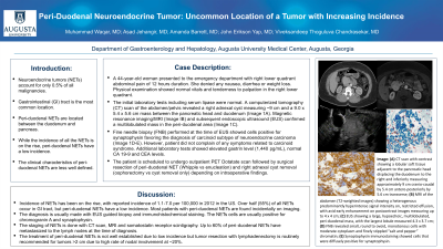
Case Description/Methods: A 44-year-old woman presented to the emergency department with right lower quadrant abdominal pain of 12 hours duration. She denied any nausea, diarrhea or weight loss. Physical examination showed normal vitals and tenderness to palpation in the right lower quadrant. The initial laboratory tests including serum lipase were normal. A computerized tomography (CT) scan of the abdomen/pelvis revealed a right adnexal cyst measuring ~9 cm and a 9.0 x 5.4 x 5.6 cm mass between the pancreatic head and duodenum (Image 1A). Magnetic resonance imaging/MRI (Image IB) and subsequent endoscopic ultrasound (EUS) confirmed a multilobulated mass in the peri-duodenal area (Image 1C). Fine needle biopsy (FNB) performed at the time of EUS showed cells positive for synaptophysin favoring the diagnosis of carcinoid subtype of neuroendocrine carcinoma (Image 1D-E). However, patient did not complain of any symptoms related to carcinoid syndrome. Additional laboratory tests showed elevated gastrin level (1,440 pg/mL), normal CA 19-9 and CEA levels . The patient is scheduled to under outpatient PET Dotatate scan followed by surgical resection of peri-duodenal NET (Whipple vs enucleation) and right adnexal cyst removal (oophorectomy vs cyst removal only) depending on intraoperative findings.
Discussion: Incidence of NETs has been on the rise, with reported incidence of 1.1-7.0 per 100,000 in 2012 in the US. Over half (55%) of all NETs occur in GI tract, but peri-duodenal NETs have a low incidence. Most patients with peri-duodenal NETs are found incidentally on imaging. The diagnosis is usually made with EUS guided biopsy and immunohistochemical staining. The NETs cells are usually positive for chromogranin A and synaptophysin. The staging of NETs is done with CT scan, MRI and somatostatin receptor scintigraphy. Up to 60% of peri-duodenal NETs have metastasized to the lymph nodes at the time of diagnosis. The treatment of peri-duodenal NETs is not well established due to low incidence but tumor resection with lymphadenectomy is routinely recommended for tumors >2 cm due to high rate of nodal involvement at ~20%.

Disclosures:
Muhammad Waqar, MD, Asad Jehangir, MD, Amanda Barrett, MD, John Erikson Yap, MD, MBA, Viveksandeep Thoguluva Chandrasekar, MD. P4144 - Peri-Duodenal Neuroendocrine Tumor: Uncommon Location of a Tumor with Increasing Incidence, ACG 2023 Annual Scientific Meeting Abstracts. Vancouver, BC, Canada: American College of Gastroenterology.
Medical College of Georgia at Augusta University, Augusta, GA
Introduction: Neuroendocrine tumors (NETs) account for only 0.5% of all malignancies and gastrointestinal (GI) tract is the most common location. Peri-duodenal NETs are located between the duodenum and pancreas. While the incidence of NETs is on the rise, peri-duodenal NETs have a low incidence and their clinical characteristics are less well defined.
Case Description/Methods: A 44-year-old woman presented to the emergency department with right lower quadrant abdominal pain of 12 hours duration. She denied any nausea, diarrhea or weight loss. Physical examination showed normal vitals and tenderness to palpation in the right lower quadrant. The initial laboratory tests including serum lipase were normal. A computerized tomography (CT) scan of the abdomen/pelvis revealed a right adnexal cyst measuring ~9 cm and a 9.0 x 5.4 x 5.6 cm mass between the pancreatic head and duodenum (Image 1A). Magnetic resonance imaging/MRI (Image IB) and subsequent endoscopic ultrasound (EUS) confirmed a multilobulated mass in the peri-duodenal area (Image 1C). Fine needle biopsy (FNB) performed at the time of EUS showed cells positive for synaptophysin favoring the diagnosis of carcinoid subtype of neuroendocrine carcinoma (Image 1D-E). However, patient did not complain of any symptoms related to carcinoid syndrome. Additional laboratory tests showed elevated gastrin level (1,440 pg/mL), normal CA 19-9 and CEA levels . The patient is scheduled to under outpatient PET Dotatate scan followed by surgical resection of peri-duodenal NET (Whipple vs enucleation) and right adnexal cyst removal (oophorectomy vs cyst removal only) depending on intraoperative findings.
Discussion: Incidence of NETs has been on the rise, with reported incidence of 1.1-7.0 per 100,000 in 2012 in the US. Over half (55%) of all NETs occur in GI tract, but peri-duodenal NETs have a low incidence. Most patients with peri-duodenal NETs are found incidentally on imaging. The diagnosis is usually made with EUS guided biopsy and immunohistochemical staining. The NETs cells are usually positive for chromogranin A and synaptophysin. The staging of NETs is done with CT scan, MRI and somatostatin receptor scintigraphy. Up to 60% of peri-duodenal NETs have metastasized to the lymph nodes at the time of diagnosis. The treatment of peri-duodenal NETs is not well established due to low incidence but tumor resection with lymphadenectomy is routinely recommended for tumors >2 cm due to high rate of nodal involvement at ~20%.

Figure: Image 1 (A): Computerized tomography of the abdomen/pelvis with intravenous contrast showing a lobular soft tissue adjacent to the pancreatic head displacing the duodenum to the right and inferiorly measuring 9.0 cm craniocaudal x 5.4 cm anteroposteriorly x 5.6 cm transverse. 1(B): Magnetic resonance imaging of the abdomen (T2-weighted images) showing a heterogenous predominantly hyperintense signal intensity on, restricted effusion, with avid early enhancement on postcontrast images measuring up to 4 x 4 cm. 1(C): Endoscopic ultrasound showing a large, hypoechoic, well-circumscribed, multilobulated, peri-duodenal mass, with the largest lobule measured 4.3 x 3.7 cm. Pressure effect of the mass on the superior mesenteric vein (SMV) noted that was displaced but no SMV invasion was noted. Portal vein, celiac axis and superior mesenteric artery appeared normal. 1 (D): 400x Diff Quik stained slide. Fine needle aspirate revealed cellular smears comprised of small, round to ovoid, monotonous cells with moderate cytoplasm and finely stippled “salt and pepper” chromatin. 1(E): 400x Synaptophyin immunostain. The cell block showed that the neoplastic cells were diffusely positive for synaptophysin.
Disclosures:
Muhammad Waqar indicated no relevant financial relationships.
Asad Jehangir indicated no relevant financial relationships.
Amanda Barrett indicated no relevant financial relationships.
John Erikson Yap indicated no relevant financial relationships.
Viveksandeep Thoguluva Chandrasekar indicated no relevant financial relationships.
Muhammad Waqar, MD, Asad Jehangir, MD, Amanda Barrett, MD, John Erikson Yap, MD, MBA, Viveksandeep Thoguluva Chandrasekar, MD. P4144 - Peri-Duodenal Neuroendocrine Tumor: Uncommon Location of a Tumor with Increasing Incidence, ACG 2023 Annual Scientific Meeting Abstracts. Vancouver, BC, Canada: American College of Gastroenterology.
