Tuesday Poster Session
Category: Colon
P3152 - Chicken Wrap in the Descending Colon: An Unusual Colonic Foreign Body
Tuesday, October 24, 2023
10:30 AM - 4:00 PM PT
Location: Exhibit Hall

Has Audio

Raj Shah, MD
University of Central Florida HCA Healthcare GME
Kissimmee, FL
Presenting Author(s)
Raj Shah, MD1, Feras Al-Moussally, MD1, Juan Oharriz, MD2
1University of Central Florida HCA Healthcare GME, Kissimmee, FL; 2Orlando VA Healthcare System, Orlando, FL
Introduction: Upper gastrointestinal tract (GIT) foreign bodies (FB) are common, with 80% occurring in children aged 6 months to 6 years. Colorectal FB, conversely, are less described in the literature as independent entities. Thus, we intend to describe a case of unusual FB in the colon and its approach.
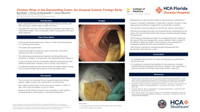
Case Description/Methods: A 54-year-old female with a history of rectal carcinoid presented for a screening colonoscopy. The patient was asymptomatic. Perianal examination showed skin tags, hemorrhoids, and normal sphincter tone with no anal tears. The colonoscopy revealed several sigmoid and descending colon diverticula. In addition, a FB was seen in the descending colon (Figure A). It was a 4x4 gauze that was successfully extracted using biopsy forceps. Additional examination revealed a 3x4 centimeter chicken bone (Figure B). On additional questioning after the procedure, the patient denied knowing how or why the foreign body ended up in her descending colon.
Discussion: The most common colorectal FB are household objects like drinking glasses, bottles, wooden rods, toothbrushes, and enema tips. Most patients present either on the same day of insertion or within 2-7 days, with a rare presentation of up to 6 months. Reasons for FB insertion include but are not limited to, self-mutilation, sexual gratification, self-treatment, or accidental swallowing.
Management of colorectal FB is based on the presence of complications. Surgery is indicated if perforation or obstruction is present. Extraction under deep general anesthesia is suggested if no complication is present. The extraction technique depends on the FB size, surface, and location. FB's above rectosigmoid junction is transabdominally manipulated into the rectum and removed trans-anally while trying to visualize potential damage by laparoscopy. If FB is large or breakable and below the rectosigmoid junction, assess for the vacuum effect and manage with a Foleys catheter. Small/sharp FB are difficult to grasp with fingers, then endoscopy and snare/basket or Rothnet is recommended for removal. After removal, checking for mucosal damage endoscopically and documenting sphincter tone is vital. It is estimated that more than 15 million colonoscopies are performed in the US each year; however, it is essential to recognize the possibility of encountering an incidental colorectal FB and be prepared with ways to navigate it, with sometimes an unusual encounter with chicken bone usually found in upper GIT.

Disclosures:
Raj Shah, MD1, Feras Al-Moussally, MD1, Juan Oharriz, MD2. P3152 - Chicken Wrap in the Descending Colon: An Unusual Colonic Foreign Body, ACG 2023 Annual Scientific Meeting Abstracts. Vancouver, BC, Canada: American College of Gastroenterology.
1University of Central Florida HCA Healthcare GME, Kissimmee, FL; 2Orlando VA Healthcare System, Orlando, FL
Introduction: Upper gastrointestinal tract (GIT) foreign bodies (FB) are common, with 80% occurring in children aged 6 months to 6 years. Colorectal FB, conversely, are less described in the literature as independent entities. Thus, we intend to describe a case of unusual FB in the colon and its approach.
Case Description/Methods: A 54-year-old female with a history of rectal carcinoid presented for a screening colonoscopy. The patient was asymptomatic. Perianal examination showed skin tags, hemorrhoids, and normal sphincter tone with no anal tears. The colonoscopy revealed several sigmoid and descending colon diverticula. In addition, a FB was seen in the descending colon (Figure A). It was a 4x4 gauze that was successfully extracted using biopsy forceps. Additional examination revealed a 3x4 centimeter chicken bone (Figure B). On additional questioning after the procedure, the patient denied knowing how or why the foreign body ended up in her descending colon.
Discussion: The most common colorectal FB are household objects like drinking glasses, bottles, wooden rods, toothbrushes, and enema tips. Most patients present either on the same day of insertion or within 2-7 days, with a rare presentation of up to 6 months. Reasons for FB insertion include but are not limited to, self-mutilation, sexual gratification, self-treatment, or accidental swallowing.
Management of colorectal FB is based on the presence of complications. Surgery is indicated if perforation or obstruction is present. Extraction under deep general anesthesia is suggested if no complication is present. The extraction technique depends on the FB size, surface, and location. FB's above rectosigmoid junction is transabdominally manipulated into the rectum and removed trans-anally while trying to visualize potential damage by laparoscopy. If FB is large or breakable and below the rectosigmoid junction, assess for the vacuum effect and manage with a Foleys catheter. Small/sharp FB are difficult to grasp with fingers, then endoscopy and snare/basket or Rothnet is recommended for removal. After removal, checking for mucosal damage endoscopically and documenting sphincter tone is vital. It is estimated that more than 15 million colonoscopies are performed in the US each year; however, it is essential to recognize the possibility of encountering an incidental colorectal FB and be prepared with ways to navigate it, with sometimes an unusual encounter with chicken bone usually found in upper GIT.

Figure: A. Colonoscopy showing foreign body which appears to be a piece of gauze.
B. Piece of bone wrapped around gauze
B. Piece of bone wrapped around gauze
Disclosures:
Raj Shah indicated no relevant financial relationships.
Feras Al-Moussally indicated no relevant financial relationships.
Juan Oharriz indicated no relevant financial relationships.
Raj Shah, MD1, Feras Al-Moussally, MD1, Juan Oharriz, MD2. P3152 - Chicken Wrap in the Descending Colon: An Unusual Colonic Foreign Body, ACG 2023 Annual Scientific Meeting Abstracts. Vancouver, BC, Canada: American College of Gastroenterology.
