Tuesday Poster Session
Category: Stomach
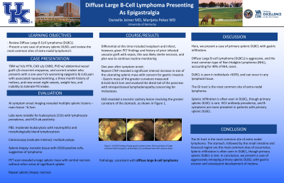
P4228 - Diffuse Large B-Cell Lymphoma Presenting as Epigastralgia
Tuesday, October 24, 2023
10:30 AM - 4:00 PM PT
Location: Exhibit Hall

Has Audio
- DJ
Danielle Joiner, MD
University of Kentucky
Lexington, KY
Presenting Author(s)
Danielle Joiner, MD, Marijeta Pekez, MD
University of Kentucky, Lexington, KY
Introduction: Diffuse Large B-Cell Lymphoma (DLBCL) is aggressive, and the most common type of Non-Hodgkin Lymphoma (NHL), accounting for 40% of NHL cases. DLBCL is seen in individuals >50YO, and can occur in any lymphoid tissue. The GI tract is the most common site of extra-nodal lymphoma. Splenic infiltration is often seen in DLBCL, though primary splenic DLBCL is rare. HCV antibody prevalence, and B-symptoms are more prevalent in patients with primary splenic DLBCL. Here we present a case of primary splenic DLBCL with gastric infiltration.
Case Description/Methods: 70M with h/o HTN, HLD, CAD s/p CABG, PVD with abdominal vessel graft c/b recurrent empyema, current smoker who presents with a one-year h/o worsening epigastric and LUQ pain, nausea/vomiting, a three-month history of melena, and new night sweats, weight loss, and inability to tolerate PO intake. At symptom onset, imaging revealed multiple splenic lesions, the main lesion measuring 6.5cm. Labs were notable for leukocytosis (21k) with lymphocyte prevalence, and HCV-ab positivity. PBS revealed moderate leukocytosis with neutrophilia and morphologically bland lymphocytosis. Colonoscopy performed for evaluation of melena revealed multiple polyps. Splenic biopsy revealed necrotic tissue with CD20 positive cells, suggestive of lymphoma. PET scan revealed a large splenic mass with central necrosis, without other areas of significant uptake. Repeat splenic biopsy revealed necrosis. DDx included neoplasm and infarct, however, given PET findings and h/o prior infected vascular graft with sepsis, likely sterile necrosis, thus plan for routine monitoring. One-year after symptom onset, repeat CTAP revealed significant interval increase in size of ulcerating splenic mass with concern for gastric invasion. Gastric mass of the greater curvature measured 8.5x10.4x13.5cm, and involved the distal tail of the pancreas with retroperitoneal lymphadenopathy concerning for metastases. EGD revealed a necrotic cavitary lesion involving the greater curvature of the stomach, as shown in figure 1. Pathology was consistent with diffuse large B-cell lymphoma.
Discussion: The GI tract is the most common site of extra-nodal lymphoma. The stomach, followed by the small intestine and ileocecal region are the most common sites of occurrence. Splenic infiltration is often seen in DLBCL, though primary splenic DLBCL is rare. In conclusion, we present a case of aggressively enlarging primary splenic DLBCL with gastric erosion and subsequent development of melena.

Disclosures:
Danielle Joiner, MD, Marijeta Pekez, MD. P4228 - Diffuse Large B-Cell Lymphoma Presenting as Epigastralgia, ACG 2023 Annual Scientific Meeting Abstracts. Vancouver, BC, Canada: American College of Gastroenterology.
University of Kentucky, Lexington, KY
Introduction: Diffuse Large B-Cell Lymphoma (DLBCL) is aggressive, and the most common type of Non-Hodgkin Lymphoma (NHL), accounting for 40% of NHL cases. DLBCL is seen in individuals >50YO, and can occur in any lymphoid tissue. The GI tract is the most common site of extra-nodal lymphoma. Splenic infiltration is often seen in DLBCL, though primary splenic DLBCL is rare. HCV antibody prevalence, and B-symptoms are more prevalent in patients with primary splenic DLBCL. Here we present a case of primary splenic DLBCL with gastric infiltration.
Case Description/Methods: 70M with h/o HTN, HLD, CAD s/p CABG, PVD with abdominal vessel graft c/b recurrent empyema, current smoker who presents with a one-year h/o worsening epigastric and LUQ pain, nausea/vomiting, a three-month history of melena, and new night sweats, weight loss, and inability to tolerate PO intake. At symptom onset, imaging revealed multiple splenic lesions, the main lesion measuring 6.5cm. Labs were notable for leukocytosis (21k) with lymphocyte prevalence, and HCV-ab positivity. PBS revealed moderate leukocytosis with neutrophilia and morphologically bland lymphocytosis. Colonoscopy performed for evaluation of melena revealed multiple polyps. Splenic biopsy revealed necrotic tissue with CD20 positive cells, suggestive of lymphoma. PET scan revealed a large splenic mass with central necrosis, without other areas of significant uptake. Repeat splenic biopsy revealed necrosis. DDx included neoplasm and infarct, however, given PET findings and h/o prior infected vascular graft with sepsis, likely sterile necrosis, thus plan for routine monitoring. One-year after symptom onset, repeat CTAP revealed significant interval increase in size of ulcerating splenic mass with concern for gastric invasion. Gastric mass of the greater curvature measured 8.5x10.4x13.5cm, and involved the distal tail of the pancreas with retroperitoneal lymphadenopathy concerning for metastases. EGD revealed a necrotic cavitary lesion involving the greater curvature of the stomach, as shown in figure 1. Pathology was consistent with diffuse large B-cell lymphoma.
Discussion: The GI tract is the most common site of extra-nodal lymphoma. The stomach, followed by the small intestine and ileocecal region are the most common sites of occurrence. Splenic infiltration is often seen in DLBCL, though primary splenic DLBCL is rare. In conclusion, we present a case of aggressively enlarging primary splenic DLBCL with gastric erosion and subsequent development of melena.

Figure: Figure 1. (A) EGD finding of large gastric cavitary lesion, (B) visualization of large cavitation (left) and gastric body (right), (C) retroflexed view with cavitary lesion.
Disclosures:
Danielle Joiner indicated no relevant financial relationships.
Marijeta Pekez indicated no relevant financial relationships.
Danielle Joiner, MD, Marijeta Pekez, MD. P4228 - Diffuse Large B-Cell Lymphoma Presenting as Epigastralgia, ACG 2023 Annual Scientific Meeting Abstracts. Vancouver, BC, Canada: American College of Gastroenterology.
