Tuesday Poster Session
Category: Stomach
P4243 - Serrated Adenoma of the Stomach: An Unusual Culprit for Potential Malignancy
Tuesday, October 24, 2023
10:30 AM - 4:00 PM PT
Location: Exhibit Hall

Has Audio

Ahmed H. abdelwahed, MBBCh
University of Connecticut-Hartford
Farmington, CT
Presenting Author(s)
Ahmed H. abdelwahed, MBBCh1, Minh Thu T. Nguyen, MD2, Steven Goldenberg, MD2
1University of Connecticut-Hartford, Farmington, CT; 2UConn Health, Farmington, CT
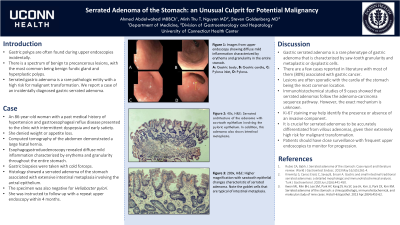
Introduction: Gastric polyps are often found during upper endoscopies incidentally. There is a spectrum of benign to precancerous lesions, with the most common being benign fundic gland and hyperplastic polyps. Serrated gastric adenoma is a rare pathologic entity with a high risk for malignant transformation. We report a case of an incidentally diagnosed gastric serrated adenoma. With very few cases reported in literature and a high proclivity towards cancer progression, our case highlights awareness of this atypical tumor.
Case Description/Methods: An 86 year-old woman with a past medical history of hypertension and gastroesophageal reflux disease presented to clinic with intermittent dyspepsia and early satiety. She denied weight or appetite loss. Computed tomography of the abdomen demonstrated a large hiatal hernia. Esophagogastroduodenoscopy revealed diffuse mild inflammation characterized by erythema and granularity throughout the entire stomach. Gastric biopsies were taken with cold forceps. Histology showed serrated adenoma of the stomach associated with extensive intestinal metaplasia involving the antral epithelium. The specimen was also negative for Heliobacter pylori. She was instructed to follow-up with a repeat upper endoscopy within 4 months.
Discussion: Gastric serrated adenoma is a rare phenotype of gastric adenoma that is characterized by saw-tooth granularity and metaplastic or dysplastic cells. There are a few cases reported in literature with a majority of them (80%) associated with gastric cancer. Lesions are often sporadic with the cardia of the stomach being the most common location. Immunohistochemical studies of 9 cases showed that serrated adenomas follow the adenoma-carcinoma sequence pathway. However, exact mechanisms are not yet identified. Ki‑67 staining may help identify the presence or absence of an invasive component. it is crucial for serrated adenomas to be accurately differentiated from villous adenoma, given its extremely high risk for malignant transformation. Patients should have close surveillance with frequent upper endoscopies to monitor for progression.

Disclosures:
Ahmed H. abdelwahed, MBBCh1, Minh Thu T. Nguyen, MD2, Steven Goldenberg, MD2. P4243 - Serrated Adenoma of the Stomach: An Unusual Culprit for Potential Malignancy, ACG 2023 Annual Scientific Meeting Abstracts. Vancouver, BC, Canada: American College of Gastroenterology.
1University of Connecticut-Hartford, Farmington, CT; 2UConn Health, Farmington, CT
Introduction: Gastric polyps are often found during upper endoscopies incidentally. There is a spectrum of benign to precancerous lesions, with the most common being benign fundic gland and hyperplastic polyps. Serrated gastric adenoma is a rare pathologic entity with a high risk for malignant transformation. We report a case of an incidentally diagnosed gastric serrated adenoma. With very few cases reported in literature and a high proclivity towards cancer progression, our case highlights awareness of this atypical tumor.
Case Description/Methods: An 86 year-old woman with a past medical history of hypertension and gastroesophageal reflux disease presented to clinic with intermittent dyspepsia and early satiety. She denied weight or appetite loss. Computed tomography of the abdomen demonstrated a large hiatal hernia. Esophagogastroduodenoscopy revealed diffuse mild inflammation characterized by erythema and granularity throughout the entire stomach. Gastric biopsies were taken with cold forceps. Histology showed serrated adenoma of the stomach associated with extensive intestinal metaplasia involving the antral epithelium. The specimen was also negative for Heliobacter pylori. She was instructed to follow-up with a repeat upper endoscopy within 4 months.
Discussion: Gastric serrated adenoma is a rare phenotype of gastric adenoma that is characterized by saw-tooth granularity and metaplastic or dysplastic cells. There are a few cases reported in literature with a majority of them (80%) associated with gastric cancer. Lesions are often sporadic with the cardia of the stomach being the most common location. Immunohistochemical studies of 9 cases showed that serrated adenomas follow the adenoma-carcinoma sequence pathway. However, exact mechanisms are not yet identified. Ki‑67 staining may help identify the presence or absence of an invasive component. it is crucial for serrated adenomas to be accurately differentiated from villous adenoma, given its extremely high risk for malignant transformation. Patients should have close surveillance with frequent upper endoscopies to monitor for progression.

Figure:
Figure (1): Images from upper endoscopy showing diffuse mild inflammation characterized by erythema and granularity in the entire stomach. A: Gastric body B:Gastric Cardia C: Pylorus inlet D: Pylrous
Figure (1): Images from upper endoscopy showing diffuse mild inflammation characterized by erythema and granularity in the entire stomach. A: Gastric body B:Gastric Cardia C: Pylorus inlet D: Pylrous
Disclosures:
Ahmed abdelwahed indicated no relevant financial relationships.
Minh Thu Nguyen indicated no relevant financial relationships.
Steven Goldenberg indicated no relevant financial relationships.
Ahmed H. abdelwahed, MBBCh1, Minh Thu T. Nguyen, MD2, Steven Goldenberg, MD2. P4243 - Serrated Adenoma of the Stomach: An Unusual Culprit for Potential Malignancy, ACG 2023 Annual Scientific Meeting Abstracts. Vancouver, BC, Canada: American College of Gastroenterology.
