Tuesday Poster Session
Category: Stomach
P4253 - Gravity Strikes Again: A Complex Gastric GIST Presenting as a Pelvic Mass
Tuesday, October 24, 2023
10:30 AM - 4:00 PM PT
Location: Exhibit Hall

Has Audio
- MM
Muhammad Mansoor, MD
Walter Reed National Military Medical Center
Bethesda, MD
Presenting Author(s)
Muhammad Mansoor, MD, Clemence White, DO, Patrick Young, MD, FACG
Walter Reed National Military Medical Center, Bethesda, MD
Introduction: The differential diagnosis for pelvic masses is broad ranging from gynecologic pathology to lower GI tract cancers. Upper GI tract pathology is rarely a consideration. We present a case of a large gastric gastrointestinal stromal tumor (GIST) presenting as a pelvic mass requiring multi-disciplinary collaboration for diagnosis and management
Case Description/Methods: A 52-year-old woman presented to the emergency department for two months of worsening pelvic pain and abdominal distention. Pelvic ultrasound demonstrated a large (13.8 x 6.3 x16 cm), complex, cystic mass in the pelvis that appeared to arrive from the left ovary. Laboratory studies revealed an elevated CA* 125 (43.2 U/mL) and CEA 19-9 (47 U/mL). She was offered pelvic mass debulking. To assist with staging, computed topographic (CT) imaging and endoscopic ultrasound (EUS) were performed prior to her scheduled surgery.
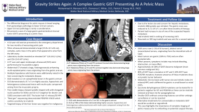
Abdominal CT showed a large, heterogeneously enhancing multi-septated pelvic mass originating from the gastric antrum. Multiple hypodense solid lesions noted in the liver concerning for metastatic disease. Esophagogastroduodenoscopy (EGD) revealed a 2 cm subepithelial lesion in the gastric antrum and EUS showed a 9.7 x 5 cm highly vascular, hypoechoic and heterogenous submucosal lesion with multi-cystic component arising from the muscularis propria. Fine needle biopsy demonstrated spindled cells with elongated nuclei positive for DOG1 and CD 117 (negative for desmin and S100) consistent with GIST. Targeted biopsy of the liver lesion was negative for malignant cells.
Due to the lesion size and concern for hepatic metastases, imatinib 400mg daily was initiated. The gastric mass was reduced to 3.2 x 4.9 x7.1 cm after three months of therapy. At the time of this report, the patient continues chemotherapy to optimize her surgical candidacy. She is also planned for repeat targeted hepatic lesion biopsy.
Discussion: GISTs are a rare (< 1% of GI tumors), distinct set of mesenchymal tumors, originating from the interstitial cells of Cajal. Most tumors are gastric (50% to 75%) and are often asymptomatic. When present, symptoms include may include bleeding, obstruction, or mass effect as in our case. This case highlights the importance of complete imaging of pelvic masses prior to embarking on surgical therapy.

Disclosures:
Muhammad Mansoor, MD, Clemence White, DO, Patrick Young, MD, FACG. P4253 - Gravity Strikes Again: A Complex Gastric GIST Presenting as a Pelvic Mass, ACG 2023 Annual Scientific Meeting Abstracts. Vancouver, BC, Canada: American College of Gastroenterology.
Walter Reed National Military Medical Center, Bethesda, MD
Introduction: The differential diagnosis for pelvic masses is broad ranging from gynecologic pathology to lower GI tract cancers. Upper GI tract pathology is rarely a consideration. We present a case of a large gastric gastrointestinal stromal tumor (GIST) presenting as a pelvic mass requiring multi-disciplinary collaboration for diagnosis and management
Case Description/Methods: A 52-year-old woman presented to the emergency department for two months of worsening pelvic pain and abdominal distention. Pelvic ultrasound demonstrated a large (13.8 x 6.3 x16 cm), complex, cystic mass in the pelvis that appeared to arrive from the left ovary. Laboratory studies revealed an elevated CA* 125 (43.2 U/mL) and CEA 19-9 (47 U/mL). She was offered pelvic mass debulking. To assist with staging, computed topographic (CT) imaging and endoscopic ultrasound (EUS) were performed prior to her scheduled surgery.
Abdominal CT showed a large, heterogeneously enhancing multi-septated pelvic mass originating from the gastric antrum. Multiple hypodense solid lesions noted in the liver concerning for metastatic disease. Esophagogastroduodenoscopy (EGD) revealed a 2 cm subepithelial lesion in the gastric antrum and EUS showed a 9.7 x 5 cm highly vascular, hypoechoic and heterogenous submucosal lesion with multi-cystic component arising from the muscularis propria. Fine needle biopsy demonstrated spindled cells with elongated nuclei positive for DOG1 and CD 117 (negative for desmin and S100) consistent with GIST. Targeted biopsy of the liver lesion was negative for malignant cells.
Due to the lesion size and concern for hepatic metastases, imatinib 400mg daily was initiated. The gastric mass was reduced to 3.2 x 4.9 x7.1 cm after three months of therapy. At the time of this report, the patient continues chemotherapy to optimize her surgical candidacy. She is also planned for repeat targeted hepatic lesion biopsy.
Discussion: GISTs are a rare (< 1% of GI tumors), distinct set of mesenchymal tumors, originating from the interstitial cells of Cajal. Most tumors are gastric (50% to 75%) and are often asymptomatic. When present, symptoms include may include bleeding, obstruction, or mass effect as in our case. This case highlights the importance of complete imaging of pelvic masses prior to embarking on surgical therapy.

Figure: A. Coronal view of the 13.8 x 6.3 x16 cm gastric mass
B. Saggital view of the 13.8 x 6.3 x16 cm gastric mass
C. Endoscopic view of 2 cm antral sub-epithelial lesion
D. EUS w/ FNB of the lesion
B. Saggital view of the 13.8 x 6.3 x16 cm gastric mass
C. Endoscopic view of 2 cm antral sub-epithelial lesion
D. EUS w/ FNB of the lesion
Disclosures:
Muhammad Mansoor: Pfizer – Stock-publicly held company(excluding mutual/index funds). Regeneron – Stock-publicly held company(excluding mutual/index funds).
Clemence White indicated no relevant financial relationships.
Patrick Young indicated no relevant financial relationships.
Muhammad Mansoor, MD, Clemence White, DO, Patrick Young, MD, FACG. P4253 - Gravity Strikes Again: A Complex Gastric GIST Presenting as a Pelvic Mass, ACG 2023 Annual Scientific Meeting Abstracts. Vancouver, BC, Canada: American College of Gastroenterology.
