Tuesday Poster Session
Category: Esophagus
P3319 - Unveiling the Uncommon: Lichenoid Esophagitis Secondary to Steven Johnson Syndrome
Tuesday, October 24, 2023
10:30 AM - 4:00 PM PT
Location: Exhibit Hall

Has Audio

Noor Hassan, MD
University of Missouri-Kansas City
Kansas City, MO
Presenting Author(s)
Islam Mohamed, MD1, Noor Hassan, MD1, Rawan Rajab, MD2, Manal Hassan, MS3, Maya Mahmoud, MD4, Khaled Elfert, MD5, Hassan Ghoz, MD1
1University of Missouri-Kansas City, Kansas City, MO; 2University of Missouri Kansas City, Kansas City, MO; 3University of Kansas, Kansas city, MO; 4Saint Louis University, St. Louis, MO; 5SBH Health System, Bronx, NY
Introduction: Lichenoid esophagitis (LE) is a rare pathology characterized by inflammation of the esophagus resembling a lichenoid pattern. The diagnosis of LE is often challenging due to its nonspecific clinical presentation and lack of a definitive histological diagnosis. In this report, we describe the case of a 47-year-old female who developed LE due to Steven Johnson syndrome (SJS) as a result of sulfasalazine (SSZ) therapy.
Case Description/Methods: A 47-year-old woman with a history of SLE presented to the GI clinic with dysphagia. Her rheumatologist had referred her after she complained of difficulty swallowing pills. Swallow evaluation showed moderate pharyngeal-esophageal dysphagia and a flexible laryngoscopy was unremarkable. EGD showed an esophageal ring. Pathology of biopsies revealed acute esophagitis with scattered fungal elements, and fluconazole was prescribed. Soon after due to poor tolerance to hydroxychloroquine and methotrexate, her rheumatologist started her on SSZ. Three weeks after, the patient presented with blistering of her lips and mucous membranes, as well as a generalized painful diffuse macular erythematous rash, indicative of SJS. The patient was intubated and placed on mechanical ventilation due to respiratory failure but was later taken off the ventilator. Several months after recovery, the patient returned to the GI clinic, this time reporting worsening dysphagia. EGD demonstrated a severe, benign-appearing intrinsic stenosis that was dilated. The patient was maintained on PPI therapy and returned for repeat EGD 4 weeks later for re-dilation and biopsies. Biopsies were sent to an outside facility that confirmed LE. The patient has undergone 5 EGDs with dilations following initial diagnosis. Despite being on various H2 blockers and PPIs, the patient's dysphagia showed minimal improvement. During the last 2 EGDs, intralesional corticosteroid injections in addition to oral fluticasone were trailed which the patient responded to well and showed improvement of her dysphagia.
Discussion: Due to the lack of previous reports of LE secondary to SJS or SSZ, the case presented highlights the importance of considering LE in the differential diagnosis of dysphagia in patients with a history of drug-induced reactions. Our experience highlights that intra-lesional steroids in addition to topical steroids can be effective for relief of dysphagia symptoms in LE. Further research is needed to better understand the pathogenesis and optimal treatment of this rare condition.

Disclosures:
Islam Mohamed, MD1, Noor Hassan, MD1, Rawan Rajab, MD2, Manal Hassan, MS3, Maya Mahmoud, MD4, Khaled Elfert, MD5, Hassan Ghoz, MD1. P3319 - Unveiling the Uncommon: Lichenoid Esophagitis Secondary to Steven Johnson Syndrome, ACG 2023 Annual Scientific Meeting Abstracts. Vancouver, BC, Canada: American College of Gastroenterology.
1University of Missouri-Kansas City, Kansas City, MO; 2University of Missouri Kansas City, Kansas City, MO; 3University of Kansas, Kansas city, MO; 4Saint Louis University, St. Louis, MO; 5SBH Health System, Bronx, NY
Introduction: Lichenoid esophagitis (LE) is a rare pathology characterized by inflammation of the esophagus resembling a lichenoid pattern. The diagnosis of LE is often challenging due to its nonspecific clinical presentation and lack of a definitive histological diagnosis. In this report, we describe the case of a 47-year-old female who developed LE due to Steven Johnson syndrome (SJS) as a result of sulfasalazine (SSZ) therapy.
Case Description/Methods: A 47-year-old woman with a history of SLE presented to the GI clinic with dysphagia. Her rheumatologist had referred her after she complained of difficulty swallowing pills. Swallow evaluation showed moderate pharyngeal-esophageal dysphagia and a flexible laryngoscopy was unremarkable. EGD showed an esophageal ring. Pathology of biopsies revealed acute esophagitis with scattered fungal elements, and fluconazole was prescribed. Soon after due to poor tolerance to hydroxychloroquine and methotrexate, her rheumatologist started her on SSZ. Three weeks after, the patient presented with blistering of her lips and mucous membranes, as well as a generalized painful diffuse macular erythematous rash, indicative of SJS. The patient was intubated and placed on mechanical ventilation due to respiratory failure but was later taken off the ventilator. Several months after recovery, the patient returned to the GI clinic, this time reporting worsening dysphagia. EGD demonstrated a severe, benign-appearing intrinsic stenosis that was dilated. The patient was maintained on PPI therapy and returned for repeat EGD 4 weeks later for re-dilation and biopsies. Biopsies were sent to an outside facility that confirmed LE. The patient has undergone 5 EGDs with dilations following initial diagnosis. Despite being on various H2 blockers and PPIs, the patient's dysphagia showed minimal improvement. During the last 2 EGDs, intralesional corticosteroid injections in addition to oral fluticasone were trailed which the patient responded to well and showed improvement of her dysphagia.
Discussion: Due to the lack of previous reports of LE secondary to SJS or SSZ, the case presented highlights the importance of considering LE in the differential diagnosis of dysphagia in patients with a history of drug-induced reactions. Our experience highlights that intra-lesional steroids in addition to topical steroids can be effective for relief of dysphagia symptoms in LE. Further research is needed to better understand the pathogenesis and optimal treatment of this rare condition.

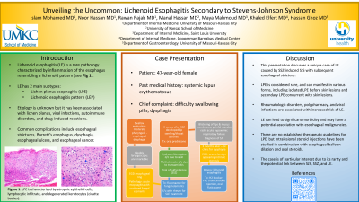
Figure: Fig illustrates acute esophagitis and a severe benign intrinsic stenosis that measures 1 cm in length.
Disclosures:
Islam Mohamed indicated no relevant financial relationships.
Noor Hassan indicated no relevant financial relationships.
Rawan Rajab indicated no relevant financial relationships.
Manal Hassan indicated no relevant financial relationships.
Maya Mahmoud indicated no relevant financial relationships.
Khaled Elfert indicated no relevant financial relationships.
Hassan Ghoz indicated no relevant financial relationships.
Islam Mohamed, MD1, Noor Hassan, MD1, Rawan Rajab, MD2, Manal Hassan, MS3, Maya Mahmoud, MD4, Khaled Elfert, MD5, Hassan Ghoz, MD1. P3319 - Unveiling the Uncommon: Lichenoid Esophagitis Secondary to Steven Johnson Syndrome, ACG 2023 Annual Scientific Meeting Abstracts. Vancouver, BC, Canada: American College of Gastroenterology.
