Tuesday Poster Session
Category: Esophagus
P3358 - A Rare Presentation of Eosinophilic-Appearing Lymphocytic Esophagitis
Tuesday, October 24, 2023
10:30 AM - 4:00 PM PT
Location: Exhibit Hall

Has Audio

Khalid Ahmed, MD
The Wright Center for GME
Scranton, PA
Presenting Author(s)
Khalid Ahmed, MD1, Udit Asija, MD1, Peter Iskander, MD2, Elmkdad Mohammed, MD1, Osama Hamid, MD, MRCPI3, Manasik Abdu, MD4, Syed Muhammad Hussain Zaidi, MD5, Abhinav Goyal, MD6
1The Wright Center for GME, Scranton, PA; 2The Wright Center for GME, Dunmore, PA; 3Cleveland Clinic, Cleveland, OH; 4University at Buffalo-Catholic Health System, Buffalo, NY; 5Wright Center, Scranton, PA; 6Geisinger Community Medical Center, Scranton, PA
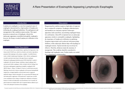
Introduction: Lymphocytic esophagitis is a rare but recognized cause of esophagitis characterized by a high number of lymphocytes infiltrating the esophageal epithelium. The pathogenesis and management of this condition remain unclear. This report presents an unusual case of dysphagia, wherein the endoscopic appearance resembled eosinophilic esophagitis; however, the biopsy revealed lymphocytic infiltration in the esophagus.
Case Description/Methods: A 67-year-old male with a medical history significant for degenerative disc disease (daily NSAID use), alcohol use disorder, and nicotine dependence presented with dysphagia as the primary complaint. A modified barium study excluded oropharyngeal dysphagia, while a barium esophagogram revealed a persistent narrowing in the upper thoracic esophagus. Subsequent esophagogastroduodenoscopy (EGD) identified LA grade A esophagitis with mucosal changes, including a ringed esophagus and a stricture/small caliber esophagus, which correlated with the findings from the barium esophagogram suggesting eosinophilic esophagitis (EoE-EREFS Edema 1, Rings 2, Exudates 0, Furrows 0, and Strictures present). Pathology showed active chronic esophagitis with increased intraepithelial lymphocytes, elongated papillae, and associated reactive squamous epithelial changes without eosinophils. He was prescribed PPI therapy and oral budesonide in applesauce. EGD performed 3 months later revealed severe candidiasis and no change in lymphocytic infiltration on biopsies, because of which oral budesonide was discontinued. Over 6 months, the patient underwent multiple EGDs with 4 sequential balloon dilations up to 11mm with subsequent resolution of his dysphagia.
Discussion: Diagnosing this condition requires a high index of suspicion due to nonspecific symptoms. Dysphagia, abdominal pain, and heartburn are commonly reported. Endoscopic appearance lacks specificity, necessitating esophageal biopsy for confirmation. About 40% of patients have an endoscopic appearance similar to eosinophilic esophagitis, highlighting the importance of lymphocytic infiltration on pathology. Symptom improvement can be achieved with proton pump inhibitors, while endoscopic dilation helps with dysphagia or esophageal stenosis. Topical steroids may not always be effective. Therefore, dilations remain the mainstay of management. This condition should be considered in dysphagia and esophagitis cases. Further studies are needed for a comprehensive understanding.

Disclosures:
Khalid Ahmed, MD1, Udit Asija, MD1, Peter Iskander, MD2, Elmkdad Mohammed, MD1, Osama Hamid, MD, MRCPI3, Manasik Abdu, MD4, Syed Muhammad Hussain Zaidi, MD5, Abhinav Goyal, MD6. P3358 - A Rare Presentation of Eosinophilic-Appearing Lymphocytic Esophagitis, ACG 2023 Annual Scientific Meeting Abstracts. Vancouver, BC, Canada: American College of Gastroenterology.
1The Wright Center for GME, Scranton, PA; 2The Wright Center for GME, Dunmore, PA; 3Cleveland Clinic, Cleveland, OH; 4University at Buffalo-Catholic Health System, Buffalo, NY; 5Wright Center, Scranton, PA; 6Geisinger Community Medical Center, Scranton, PA
Introduction: Lymphocytic esophagitis is a rare but recognized cause of esophagitis characterized by a high number of lymphocytes infiltrating the esophageal epithelium. The pathogenesis and management of this condition remain unclear. This report presents an unusual case of dysphagia, wherein the endoscopic appearance resembled eosinophilic esophagitis; however, the biopsy revealed lymphocytic infiltration in the esophagus.
Case Description/Methods: A 67-year-old male with a medical history significant for degenerative disc disease (daily NSAID use), alcohol use disorder, and nicotine dependence presented with dysphagia as the primary complaint. A modified barium study excluded oropharyngeal dysphagia, while a barium esophagogram revealed a persistent narrowing in the upper thoracic esophagus. Subsequent esophagogastroduodenoscopy (EGD) identified LA grade A esophagitis with mucosal changes, including a ringed esophagus and a stricture/small caliber esophagus, which correlated with the findings from the barium esophagogram suggesting eosinophilic esophagitis (EoE-EREFS Edema 1, Rings 2, Exudates 0, Furrows 0, and Strictures present). Pathology showed active chronic esophagitis with increased intraepithelial lymphocytes, elongated papillae, and associated reactive squamous epithelial changes without eosinophils. He was prescribed PPI therapy and oral budesonide in applesauce. EGD performed 3 months later revealed severe candidiasis and no change in lymphocytic infiltration on biopsies, because of which oral budesonide was discontinued. Over 6 months, the patient underwent multiple EGDs with 4 sequential balloon dilations up to 11mm with subsequent resolution of his dysphagia.
Discussion: Diagnosing this condition requires a high index of suspicion due to nonspecific symptoms. Dysphagia, abdominal pain, and heartburn are commonly reported. Endoscopic appearance lacks specificity, necessitating esophageal biopsy for confirmation. About 40% of patients have an endoscopic appearance similar to eosinophilic esophagitis, highlighting the importance of lymphocytic infiltration on pathology. Symptom improvement can be achieved with proton pump inhibitors, while endoscopic dilation helps with dysphagia or esophageal stenosis. Topical steroids may not always be effective. Therefore, dilations remain the mainstay of management. This condition should be considered in dysphagia and esophagitis cases. Further studies are needed for a comprehensive understanding.

Figure: Figure 1. A) Upper third of esophagus showing eosinophilic esophagitis and stenosis. B) Middle third of esophagus showing eosinophilic esophagitis.
Disclosures:
Khalid Ahmed indicated no relevant financial relationships.
Udit Asija indicated no relevant financial relationships.
Peter Iskander indicated no relevant financial relationships.
Elmkdad Mohammed indicated no relevant financial relationships.
Osama Hamid indicated no relevant financial relationships.
Manasik Abdu indicated no relevant financial relationships.
Syed Muhammad Hussain Zaidi indicated no relevant financial relationships.
Abhinav Goyal: Moderna – Stock-publicly held company(excluding mutual/index funds). Pfiezer – Stock-publicly held company(excluding mutual/index funds).
Khalid Ahmed, MD1, Udit Asija, MD1, Peter Iskander, MD2, Elmkdad Mohammed, MD1, Osama Hamid, MD, MRCPI3, Manasik Abdu, MD4, Syed Muhammad Hussain Zaidi, MD5, Abhinav Goyal, MD6. P3358 - A Rare Presentation of Eosinophilic-Appearing Lymphocytic Esophagitis, ACG 2023 Annual Scientific Meeting Abstracts. Vancouver, BC, Canada: American College of Gastroenterology.
