Monday Poster Session
Category: Biliary/Pancreas
P1485 - Duodenal Stenosis from Groove Pancreatitis; A Rare Phenomenon
Monday, October 23, 2023
10:30 AM - 4:15 PM PT
Location: Exhibit Hall

Has Audio

Mounir Ibrahim, MD
Cleveland Clinic Foundation
Cleveland, OH
Presenting Author(s)
Somtochukwu Onwuzo, MD1, Mounir Ibrahim, MD2, Chidera Onwuzo, MBBS3, Antoine Boustany, MD2, Wassan Fawzi. Safadi, MD4, Maryam Hijjawai, MD4, Mohammed Janajreh, MD5, Noor Eddin Abdel Jalil, MD4, Ala Abdel-Jalil, MD2
1Cleveland Clinic Foundation, Fairview Park, OH; 2Cleveland Clinic Foundation, Cleveland, OH; 3General Hospital Lagos, Fairview Park, OH; 4University of Jordan, Cleveland, OH; 5Al-Najah University, Cleveland, OH
Introduction: Groove pancreatitis (GP) is a rare form of chronic pancreatitis affecting the pancreaticoduodenal groove, a narrow space between the pancreatic head, duodenum, and common bile duct. It is usually seen in male patients who consume alcohol. This condition is frequently under-recognized, which causes treatment delay and possible serious complications. We present a case of idiopathic groove pancreatitis in a female patient with a history of recurrent pancreatitis.
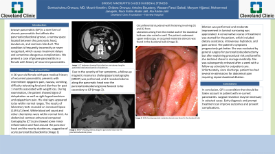
Case Description/Methods: A 56-year-old female presented with recurrent epigastric pain, nausea, vomiting, diarrhea, with 25 pounds of weight loss. The patient was a non-smoker with no history of alcohol use. On examination, she had epigastric tenderness. Laboratory tests revealed increased serum lipase level (149 U/L) but otherwise was unremarkable. Workup for other etiologies for acute & chronic pancreatitis was negative. An abdominal computed tomography (CT) scan showed inflammatory changes around the pancreatic head and the duodenal groove, suggesting acute pancreatitis & duodenitis (Image 1). A follow-up magnetic resonance cholangiopancreatography (MRCP) revealed edema along the pancreatic head near the pancreaticoduodenal groove along with intraduodenal wall cysts and circumferential duodenal wall thickening involving the bulb and second portion, favored to be secondary to Groove Pancreatitis (Image 2). Upper endoscopy which showed acquired moderate stenosis in the duodenal bulb (Image 3). Endosonograophy was done to delineate further the anatomy, which showed significant duodenal wall thickening and intraduodenal wall cystic changes, but without solid masses within the pancreatic head or duodenal wall (Image 4). A conservative treatment with dietary modification and gradual endoscopic dilation of the stricture using CRE-balloons to 15 mm (Boston Scientific, Marlborough, MA) with moderate improvement in the luminal narrowing. Patient's symptoms progressively improved. Patient was evaluated by Surgery for consideration of pancreaticoduodenectomy (Whipple procedure) for more definitive therapy, but she chose to continue to manage medically.
Discussion: Groove Pancreatitis is a rare condition, usually seen in males who use alcohol. It can result in recurrent acute or chronic pancreatitis. It can lead to malnutrition. Malignancy and other etiologies ruled out. Surgical resection might be necessary in select cases. Early diagnosis and prompt treatment are important to improve outcome and prevent further complications.

Disclosures:
Somtochukwu Onwuzo, MD1, Mounir Ibrahim, MD2, Chidera Onwuzo, MBBS3, Antoine Boustany, MD2, Wassan Fawzi. Safadi, MD4, Maryam Hijjawai, MD4, Mohammed Janajreh, MD5, Noor Eddin Abdel Jalil, MD4, Ala Abdel-Jalil, MD2. P1485 - Duodenal Stenosis from Groove Pancreatitis; A Rare Phenomenon, ACG 2023 Annual Scientific Meeting Abstracts. Vancouver, BC, Canada: American College of Gastroenterology.
1Cleveland Clinic Foundation, Fairview Park, OH; 2Cleveland Clinic Foundation, Cleveland, OH; 3General Hospital Lagos, Fairview Park, OH; 4University of Jordan, Cleveland, OH; 5Al-Najah University, Cleveland, OH
Introduction: Groove pancreatitis (GP) is a rare form of chronic pancreatitis affecting the pancreaticoduodenal groove, a narrow space between the pancreatic head, duodenum, and common bile duct. It is usually seen in male patients who consume alcohol. This condition is frequently under-recognized, which causes treatment delay and possible serious complications. We present a case of idiopathic groove pancreatitis in a female patient with a history of recurrent pancreatitis.
Case Description/Methods: A 56-year-old female presented with recurrent epigastric pain, nausea, vomiting, diarrhea, with 25 pounds of weight loss. The patient was a non-smoker with no history of alcohol use. On examination, she had epigastric tenderness. Laboratory tests revealed increased serum lipase level (149 U/L) but otherwise was unremarkable. Workup for other etiologies for acute & chronic pancreatitis was negative. An abdominal computed tomography (CT) scan showed inflammatory changes around the pancreatic head and the duodenal groove, suggesting acute pancreatitis & duodenitis (Image 1). A follow-up magnetic resonance cholangiopancreatography (MRCP) revealed edema along the pancreatic head near the pancreaticoduodenal groove along with intraduodenal wall cysts and circumferential duodenal wall thickening involving the bulb and second portion, favored to be secondary to Groove Pancreatitis (Image 2). Upper endoscopy which showed acquired moderate stenosis in the duodenal bulb (Image 3). Endosonograophy was done to delineate further the anatomy, which showed significant duodenal wall thickening and intraduodenal wall cystic changes, but without solid masses within the pancreatic head or duodenal wall (Image 4). A conservative treatment with dietary modification and gradual endoscopic dilation of the stricture using CRE-balloons to 15 mm (Boston Scientific, Marlborough, MA) with moderate improvement in the luminal narrowing. Patient's symptoms progressively improved. Patient was evaluated by Surgery for consideration of pancreaticoduodenectomy (Whipple procedure) for more definitive therapy, but she chose to continue to manage medically.
Discussion: Groove Pancreatitis is a rare condition, usually seen in males who use alcohol. It can result in recurrent acute or chronic pancreatitis. It can lead to malnutrition. Malignancy and other etiologies ruled out. Surgical resection might be necessary in select cases. Early diagnosis and prompt treatment are important to improve outcome and prevent further complications.

Figure: CT , MRCP and EGD with EUS showing edema along the pancreatic head alongside stenosis of the duodenal bulb
Disclosures:
Somtochukwu Onwuzo indicated no relevant financial relationships.
Mounir Ibrahim indicated no relevant financial relationships.
Chidera Onwuzo indicated no relevant financial relationships.
Antoine Boustany indicated no relevant financial relationships.
Wassan Safadi indicated no relevant financial relationships.
Maryam Hijjawai indicated no relevant financial relationships.
Mohammed Janajreh indicated no relevant financial relationships.
Noor Eddin Abdel Jalil indicated no relevant financial relationships.
Ala Abdel-Jalil indicated no relevant financial relationships.
Somtochukwu Onwuzo, MD1, Mounir Ibrahim, MD2, Chidera Onwuzo, MBBS3, Antoine Boustany, MD2, Wassan Fawzi. Safadi, MD4, Maryam Hijjawai, MD4, Mohammed Janajreh, MD5, Noor Eddin Abdel Jalil, MD4, Ala Abdel-Jalil, MD2. P1485 - Duodenal Stenosis from Groove Pancreatitis; A Rare Phenomenon, ACG 2023 Annual Scientific Meeting Abstracts. Vancouver, BC, Canada: American College of Gastroenterology.
