Monday Poster Session
Category: Biliary/Pancreas
P1534 - Recurrent Cholangitis from Coil Migration After Embolization of Cystic Artery Pseudoaneurysm
Monday, October 23, 2023
10:30 AM - 4:15 PM PT
Location: Exhibit Hall

Has Audio
- KT
Kshitij Thakur, MD, MSc
University of Kentucky
Lexington, Kentucky
Presenting Author(s)
Jeeva Jaganathan, MD1, Ujas Patel, MD2, Kshitij Thakur, MD, MSc2, Samuel Mardini, MD, MBA, MPH2
1University of Kentucky School of Medicine, Lexington, KY; 2University of Kentucky, Lexington, KY
Introduction: Cystic artery pseudoaneurysm is a rare complication following laparoscopic cholecystectomy. Approximately 1/3rd can present with cholestatic jaundice and transaminitis from external compression of the bile duct in addition to abdominal pain and hemobilia. Endoscopic biliary stenting and coil embolization of the aneurysm is effective but can have rare complications. We present a case of biliary obstruction from a migrated coil causing recurrent cholangitis resulting in decompensated cirrhosis.
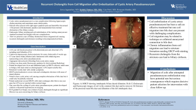
Case Description/Methods: 64 yo female presented with nausea, abdominal pain and elevated liver panel 8 weeks after an uncomplicated laparoscopic cholecystectomy. CT showed biliary dilatation and a hematoma in the gallbladder fossa with an enhancing area representing a cystic artery pseudoaneurysm. An angiogram showed active bleeding from the pseudoaneurysm which was coiled under fluoroscopy. ERCP showed fistula extravasation of contrast from mid duct which was stented. Her hospital course was complicated by a bile leak requiring repeat stenting and she was discharged with improving liver enzymes. She had relapsing symptoms of abdominal pain, nausea and abnormal liver panel. An MR/ERCP performed 8 months after showed intra and extrahepatic biliary dilatation with 2 cm cystic artery coil causing near complete obstruction of main bile duct (fig A,B,C). Coil was partially removed endoscopically. ERCP was repeated thrice demonstrating recurrent stent occlusion and thinning of the duct with unsuccessful attempts to remove the residual coil. Liver enzymes were continually elevated with ALP persistently over 500. A year later, she presented with hematemesis and confusion with imaging findings of cirrhosis and ascites. Ascitic fluid was consistent with portal hypertension. EGD showed esophageal varices. ERCP showed narrowing and irregularity of the proximal main bile duct in the area of the coil with upstream dilatation (fig D). A EUS guided liver biopsy showed acute cholangitis.
Discussion: Coil migration into the biliary system is a rare complication of vascular embolization with risk factors such as the technique and presence of collaterals. Endoscopic removal with stenting reverses clinical and biochemical abnormalities. Rarely, chronic inflammation can cause recurrent strictures and cholangitis leading to biliary cirrhosis as in our patient. Coil migration into bile duct can have serious consequences and requires proper patient selection and close follow-up.

Disclosures:
Jeeva Jaganathan, MD1, Ujas Patel, MD2, Kshitij Thakur, MD, MSc2, Samuel Mardini, MD, MBA, MPH2. P1534 - Recurrent Cholangitis from Coil Migration After Embolization of Cystic Artery Pseudoaneurysm, ACG 2023 Annual Scientific Meeting Abstracts. Vancouver, BC, Canada: American College of Gastroenterology.
1University of Kentucky School of Medicine, Lexington, KY; 2University of Kentucky, Lexington, KY
Introduction: Cystic artery pseudoaneurysm is a rare complication following laparoscopic cholecystectomy. Approximately 1/3rd can present with cholestatic jaundice and transaminitis from external compression of the bile duct in addition to abdominal pain and hemobilia. Endoscopic biliary stenting and coil embolization of the aneurysm is effective but can have rare complications. We present a case of biliary obstruction from a migrated coil causing recurrent cholangitis resulting in decompensated cirrhosis.
Case Description/Methods: 64 yo female presented with nausea, abdominal pain and elevated liver panel 8 weeks after an uncomplicated laparoscopic cholecystectomy. CT showed biliary dilatation and a hematoma in the gallbladder fossa with an enhancing area representing a cystic artery pseudoaneurysm. An angiogram showed active bleeding from the pseudoaneurysm which was coiled under fluoroscopy. ERCP showed fistula extravasation of contrast from mid duct which was stented. Her hospital course was complicated by a bile leak requiring repeat stenting and she was discharged with improving liver enzymes. She had relapsing symptoms of abdominal pain, nausea and abnormal liver panel. An MR/ERCP performed 8 months after showed intra and extrahepatic biliary dilatation with 2 cm cystic artery coil causing near complete obstruction of main bile duct (fig A,B,C). Coil was partially removed endoscopically. ERCP was repeated thrice demonstrating recurrent stent occlusion and thinning of the duct with unsuccessful attempts to remove the residual coil. Liver enzymes were continually elevated with ALP persistently over 500. A year later, she presented with hematemesis and confusion with imaging findings of cirrhosis and ascites. Ascitic fluid was consistent with portal hypertension. EGD showed esophageal varices. ERCP showed narrowing and irregularity of the proximal main bile duct in the area of the coil with upstream dilatation (fig D). A EUS guided liver biopsy showed acute cholangitis.
Discussion: Coil migration into the biliary system is a rare complication of vascular embolization with risk factors such as the technique and presence of collaterals. Endoscopic removal with stenting reverses clinical and biochemical abnormalities. Rarely, chronic inflammation can cause recurrent strictures and cholangitis leading to biliary cirrhosis as in our patient. Coil migration into bile duct can have serious consequences and requires proper patient selection and close follow-up.

Figure: Figures: A) MRCP showing intra-hepatic biliary ductal dilatation. B) Endoscopic image of coil in the common bile duct. C) Fluoroscopic coil removal. D) Stricture at the proximal main bile duct and dilatation of the left intra-hepatic duct.
Disclosures:
Jeeva Jaganathan indicated no relevant financial relationships.
Ujas Patel indicated no relevant financial relationships.
Kshitij Thakur indicated no relevant financial relationships.
Samuel Mardini indicated no relevant financial relationships.
Jeeva Jaganathan, MD1, Ujas Patel, MD2, Kshitij Thakur, MD, MSc2, Samuel Mardini, MD, MBA, MPH2. P1534 - Recurrent Cholangitis from Coil Migration After Embolization of Cystic Artery Pseudoaneurysm, ACG 2023 Annual Scientific Meeting Abstracts. Vancouver, BC, Canada: American College of Gastroenterology.
