Monday Poster Session
Category: Biliary/Pancreas
P1558 - Recurrent Acute Pancreatitis in Pregnancy - It's Not Always a Stone
Monday, October 23, 2023
10:30 AM - 4:15 PM PT
Location: Exhibit Hall

Has Audio
.jpg)
Sarah Huang, MD
New York Presbyterian Brooklyn Methodist Hospital
Brooklyn, New York
Presenting Author(s)
Sarah Huang, MD, Karthik Chandrasekaran, MD, Pratishtha Singh, MD, Arshish Dua, MD
New York Presbyterian Brooklyn Methodist Hospital, Brooklyn, NY
Introduction: Biliary pathology during pregnancy is commonly due to cholestatic entities. Ampullary adenoma during pregnancy is less reported with only 2 cases described in current literature. We report the first case of recurrent pancreatitis secondary to an ampullary adenoma with observed progression to adenocarcinoma during pregnancy.
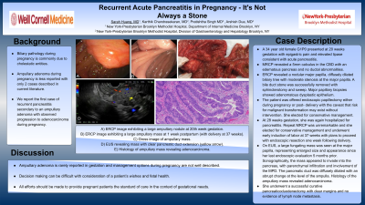
Case Description/Methods: A 34 year old female G1P0 presented at 20 weeks gestation with epigastric pain and elevated lipase consistent with acute pancreatitis. MRCP revealed a 5mm calculus in the common bile duct with an edematous pancreas and no ductal abnormalities. After multidisciplinary discussion, a decision was made to proceed with endoscopy. ERCP revealed a nodular major papilla with concern for an underlying lesion. The biliary tree was diffusely dilated with moderate stenosis at the major papilla. A bile duct stone was successfully removed with sphincterotomy and sweep. Major papillary biopsies showed adenomatous dysplastic epithelium. After multidisciplinary discussion, the patient was offered endoscopic papillectomy either during pregnancy or after her delivery with the caveat that risk for malignant transformation may exist without intervention. She elected for conservative management. She was again hospitalized at 28 weeks gestation with pancreatitis and repeat MRCP was unremarkable for any frank mass or ductal dilation. She again elected for conservative management and underwent early induction of labor at 37 weeks with plans to proceed with endoscopic resection one week following delivery. On EUS, a large fungating mass was seen at the major papilla, representing a significant change in size and appearance since her last endoscopic evaluation 5 months prior. Sonographically, the mass appeared to invade into the pancreas, with parenchymal infiltration and involvement of the main pancreatic duct. The pancreatic duct was diffusely dilated with an abrupt change at the level of the ampulla. Histology of the mass revealed adenocarcinoma. Following multidisciplinary discussion, she underwent a successful curative pancreaticoduodenectomy with clear margins and no evidence of lymph node metastasis.
Discussion: Ampullary adenoma is rarely reported in gestation and management options during pregnancy are not well described. Decision making can be difficult with consideration of a patient’s wishes and fetal health. All efforts should be made to provide pregnant patients the standard of care in the context of their gestational needs.

Disclosures:
Sarah Huang, MD, Karthik Chandrasekaran, MD, Pratishtha Singh, MD, Arshish Dua, MD. P1558 - Recurrent Acute Pancreatitis in Pregnancy - It's Not Always a Stone, ACG 2023 Annual Scientific Meeting Abstracts. Vancouver, BC, Canada: American College of Gastroenterology.
New York Presbyterian Brooklyn Methodist Hospital, Brooklyn, NY
Introduction: Biliary pathology during pregnancy is commonly due to cholestatic entities. Ampullary adenoma during pregnancy is less reported with only 2 cases described in current literature. We report the first case of recurrent pancreatitis secondary to an ampullary adenoma with observed progression to adenocarcinoma during pregnancy.
Case Description/Methods: A 34 year old female G1P0 presented at 20 weeks gestation with epigastric pain and elevated lipase consistent with acute pancreatitis. MRCP revealed a 5mm calculus in the common bile duct with an edematous pancreas and no ductal abnormalities. After multidisciplinary discussion, a decision was made to proceed with endoscopy. ERCP revealed a nodular major papilla with concern for an underlying lesion. The biliary tree was diffusely dilated with moderate stenosis at the major papilla. A bile duct stone was successfully removed with sphincterotomy and sweep. Major papillary biopsies showed adenomatous dysplastic epithelium. After multidisciplinary discussion, the patient was offered endoscopic papillectomy either during pregnancy or after her delivery with the caveat that risk for malignant transformation may exist without intervention. She elected for conservative management. She was again hospitalized at 28 weeks gestation with pancreatitis and repeat MRCP was unremarkable for any frank mass or ductal dilation. She again elected for conservative management and underwent early induction of labor at 37 weeks with plans to proceed with endoscopic resection one week following delivery. On EUS, a large fungating mass was seen at the major papilla, representing a significant change in size and appearance since her last endoscopic evaluation 5 months prior. Sonographically, the mass appeared to invade into the pancreas, with parenchymal infiltration and involvement of the main pancreatic duct. The pancreatic duct was diffusely dilated with an abrupt change at the level of the ampulla. Histology of the mass revealed adenocarcinoma. Following multidisciplinary discussion, she underwent a successful curative pancreaticoduodenectomy with clear margins and no evidence of lymph node metastasis.
Discussion: Ampullary adenoma is rarely reported in gestation and management options during pregnancy are not well described. Decision making can be difficult with consideration of a patient’s wishes and fetal health. All efforts should be made to provide pregnant patients the standard of care in the context of their gestational needs.

Figure: A) ERCP image exhibiting a large ampullary nodule at 20th week gestation.
B) ERCP image exhibiting a large ampullary mass at 1 week postpartum (with delivery at 37 weeks).
C) Gross image of ampullary mass.
D) EUS revealing mass with clear pancreatic duct extension (yellow arrow).
E) Histology of ampullary mass biopsy revealing adenocarcinoma.
B) ERCP image exhibiting a large ampullary mass at 1 week postpartum (with delivery at 37 weeks).
C) Gross image of ampullary mass.
D) EUS revealing mass with clear pancreatic duct extension (yellow arrow).
E) Histology of ampullary mass biopsy revealing adenocarcinoma.
Disclosures:
Sarah Huang indicated no relevant financial relationships.
Karthik Chandrasekaran indicated no relevant financial relationships.
Pratishtha Singh indicated no relevant financial relationships.
Arshish Dua indicated no relevant financial relationships.
Sarah Huang, MD, Karthik Chandrasekaran, MD, Pratishtha Singh, MD, Arshish Dua, MD. P1558 - Recurrent Acute Pancreatitis in Pregnancy - It's Not Always a Stone, ACG 2023 Annual Scientific Meeting Abstracts. Vancouver, BC, Canada: American College of Gastroenterology.
