Monday Poster Session
Category: Biliary/Pancreas
P1559 - Intraductal Papillary Mucinous Neoplasm Coexisting With Pancreatic Divisum
Monday, October 23, 2023
10:30 AM - 4:15 PM PT
Location: Exhibit Hall

Has Audio

Farhan Azad, DO
University at Buffalo
Buffalo, New York
Presenting Author(s)
Farhan Azad, DO, Clive J. Miranda, DO, MS, Alexander M. Carlson, DO, Prutha Patel, DO, Muddasir Ayaz, MD
University at Buffalo, Buffalo, NY
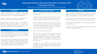
Introduction: Pancreatic divisum (PD) is a congenital malformation caused by the failure of fusion of the ventral and dorsal pancreatic duct systems. It is typically asymptomatic. However, recurrent pancreatitis with progression to chronic pancreatitis is seen in 25-38% of patients. While pancreatic tumors can occur in PD, intraductal papillary mucinous neoplasm (IPMN) is rare and has a high rate of malignant transformation, especially when seen in conjunction with main pancreatic duct (MPD) dilation.
Case Description/Methods: A 62-year-old female with a history of recurrent and chronic pancreatitis and pain as long-term sequelae presented with worsening epigastric pain, recent weight loss, and decreased appetite. She had a history of PD diagnosed 15 years prior. She also had hospitalizations at least once a year for pancreatitis, with some visits requiring supplementation with hyperalimentation. Prior surgeries included cholecystectomy and appendectomy. She had no history of alcohol use. On exam, her abdomen was tender to palpation in the epigastric area. Laboratory values revealed an elevated AST, ALT, and ALP at 58 IU/L, 91 IU/L, and 476 IU/L respectively. CEA was elevated at 18.2 ng/mL, while CA19-9 was < 3 units/mL. CT of the abdomen revealed a 4 cm cystic mass in the head of the pancreas, cystic with solid components. A markedly dilated pancreatic duct in the tail and body of the pancreas was noted. ERCP showed the pancreatic duct communicating with the mass lesion. Biopsy confirmed the mass to be IPMN. Given the size of the mass, ductal obstruction with pain, and risk for malignancy, she underwent a pyloric-preserving Whipple procedure with improved epigastric symptoms following the procedure. The goal was to dissect the mass and anastomosis to decompress the pancreatic duct. A repeat CT scan revealed stable diffuse atrophy, calcification throughout the pancreatic body and tail, and decreased gross ductal dilatation of the pancreatic duct with no new lesions.
Discussion: IPMN associated with MPD dilation accounts for 15-21% of IPMNs, typically located in the pancreatic head, and has the highest risk of malignancy. Surgery is offered in these cases, as was done here. Among reported cases of IPMN with PD, the tumor is typically seen in the dorsal pancreas, compared to the ventral pancreas. Some suggest that IPMN itself is a cause of chronic pancreatitis and not vice versa. It is difficult to elucidate the etiology between PD and IPMN, and further studies are required.
Disclosures:
Farhan Azad, DO, Clive J. Miranda, DO, MS, Alexander M. Carlson, DO, Prutha Patel, DO, Muddasir Ayaz, MD. P1559 - Intraductal Papillary Mucinous Neoplasm Coexisting With Pancreatic Divisum, ACG 2023 Annual Scientific Meeting Abstracts. Vancouver, BC, Canada: American College of Gastroenterology.
University at Buffalo, Buffalo, NY
Introduction: Pancreatic divisum (PD) is a congenital malformation caused by the failure of fusion of the ventral and dorsal pancreatic duct systems. It is typically asymptomatic. However, recurrent pancreatitis with progression to chronic pancreatitis is seen in 25-38% of patients. While pancreatic tumors can occur in PD, intraductal papillary mucinous neoplasm (IPMN) is rare and has a high rate of malignant transformation, especially when seen in conjunction with main pancreatic duct (MPD) dilation.
Case Description/Methods: A 62-year-old female with a history of recurrent and chronic pancreatitis and pain as long-term sequelae presented with worsening epigastric pain, recent weight loss, and decreased appetite. She had a history of PD diagnosed 15 years prior. She also had hospitalizations at least once a year for pancreatitis, with some visits requiring supplementation with hyperalimentation. Prior surgeries included cholecystectomy and appendectomy. She had no history of alcohol use. On exam, her abdomen was tender to palpation in the epigastric area. Laboratory values revealed an elevated AST, ALT, and ALP at 58 IU/L, 91 IU/L, and 476 IU/L respectively. CEA was elevated at 18.2 ng/mL, while CA19-9 was < 3 units/mL. CT of the abdomen revealed a 4 cm cystic mass in the head of the pancreas, cystic with solid components. A markedly dilated pancreatic duct in the tail and body of the pancreas was noted. ERCP showed the pancreatic duct communicating with the mass lesion. Biopsy confirmed the mass to be IPMN. Given the size of the mass, ductal obstruction with pain, and risk for malignancy, she underwent a pyloric-preserving Whipple procedure with improved epigastric symptoms following the procedure. The goal was to dissect the mass and anastomosis to decompress the pancreatic duct. A repeat CT scan revealed stable diffuse atrophy, calcification throughout the pancreatic body and tail, and decreased gross ductal dilatation of the pancreatic duct with no new lesions.
Discussion: IPMN associated with MPD dilation accounts for 15-21% of IPMNs, typically located in the pancreatic head, and has the highest risk of malignancy. Surgery is offered in these cases, as was done here. Among reported cases of IPMN with PD, the tumor is typically seen in the dorsal pancreas, compared to the ventral pancreas. Some suggest that IPMN itself is a cause of chronic pancreatitis and not vice versa. It is difficult to elucidate the etiology between PD and IPMN, and further studies are required.
Disclosures:
Farhan Azad indicated no relevant financial relationships.
Clive Miranda indicated no relevant financial relationships.
Alexander Carlson indicated no relevant financial relationships.
Prutha Patel indicated no relevant financial relationships.
Muddasir Ayaz indicated no relevant financial relationships.
Farhan Azad, DO, Clive J. Miranda, DO, MS, Alexander M. Carlson, DO, Prutha Patel, DO, Muddasir Ayaz, MD. P1559 - Intraductal Papillary Mucinous Neoplasm Coexisting With Pancreatic Divisum, ACG 2023 Annual Scientific Meeting Abstracts. Vancouver, BC, Canada: American College of Gastroenterology.
