Monday Poster Session
Category: Biliary/Pancreas
P1577 - A Case of Metastatic Seminoma Mimicking a Primary Pancreatic Tumor
Monday, October 23, 2023
10:30 AM - 4:15 PM PT
Location: Exhibit Hall

Has Audio
- MO
Mark Obri, MD
Henry Ford Hospital
Detroit, Michigan
Presenting Author(s)
Mark Obri, MD, William Davis, DO, Muhammad Zarrar Khan, MD, Hamna Fahad, MD, John Curran, MD, Robert Pompa, MD
Henry Ford Hospital, Detroit, MI
Introduction: Patients who present with painless jaundice, dark urine, and pruritis immediately spark concern for a primary pancreatic malignant tumor. Alternatively, the patient can present with a malignancy from an unknown origin. We present a rare case of a metastatic seminoma mimicking a primary pancreatic tumor and highlight the importance of biopsy and proper pathology.
Case Description/Methods:
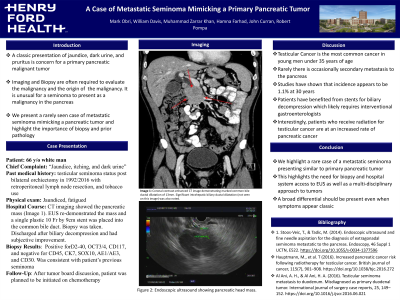
A 66 y/o male with past medical history of testicular seminoma status post bilateral orchiectomy in 1992/2016 with retroperitoneal lymph node resection who presented with a chief complaint of pruritis, jaundice, and abdominal pain for three weeks prior to presentation. His liver profile, computed tomography (CT), and Magnetic Resonance Imaging (MRI) of his abdomen showed concerns of extrinsic biliary compression with internal and external biliary dilation (Image 1A). Diagnostic imaging did not show additional areas of metastasis.
For diagnostic sampling an endoscopic ultrasound was performed which re-demonstrated the pancreatic head mass which was biopsied through fine needle aspiration (FNA). Following this, an endoscopic retrograde cholangiopancreatography was performed with placement of a single plastic 10 Fr by 9cm stent into the common bile duct (Image 1B).
FNA biopsy showed neoplastic cells positive for D2-40, OCT3/4, CD117, and negative for CD45, CK7, SOX10, AE1/AE3, and CD30 which was consistent with a seminoma that was thought to be a distant metastasis from patients primary seminoma. Prior to discharge the patient’s liver biochemistry panel was shown to downtrend after biliary decompression. Patient was planned to begin chemotherapy in the outpatient setting after tumor board discussion.
Discussion: The presenting symptoms of painless jaundice with imaging showing a pancreatic head mass raised concerns for pancreatic adenocarcinoma, however on pathologic review it was shown to be a solitary metastatic germ cell tumors. This delineation through proper sampling and histopathologic review was critical for proper diagnosis and treatment of a curable malignancy. This case is unique that patient had suspected cured testicular seminoma with now new metastasis seven years after orchiectomy and retroperitoneal lymph node resection along with the pancreas being the sole area of distant metastasis. This highlights the importance for a broad differential diagnosis and the importance of having trained gastroenterologists available for biopsies of pancreatic masses.

Disclosures:
Mark Obri, MD, William Davis, DO, Muhammad Zarrar Khan, MD, Hamna Fahad, MD, John Curran, MD, Robert Pompa, MD. P1577 - A Case of Metastatic Seminoma Mimicking a Primary Pancreatic Tumor, ACG 2023 Annual Scientific Meeting Abstracts. Vancouver, BC, Canada: American College of Gastroenterology.
Henry Ford Hospital, Detroit, MI
Introduction: Patients who present with painless jaundice, dark urine, and pruritis immediately spark concern for a primary pancreatic malignant tumor. Alternatively, the patient can present with a malignancy from an unknown origin. We present a rare case of a metastatic seminoma mimicking a primary pancreatic tumor and highlight the importance of biopsy and proper pathology.
Case Description/Methods:
A 66 y/o male with past medical history of testicular seminoma status post bilateral orchiectomy in 1992/2016 with retroperitoneal lymph node resection who presented with a chief complaint of pruritis, jaundice, and abdominal pain for three weeks prior to presentation. His liver profile, computed tomography (CT), and Magnetic Resonance Imaging (MRI) of his abdomen showed concerns of extrinsic biliary compression with internal and external biliary dilation (Image 1A). Diagnostic imaging did not show additional areas of metastasis.
For diagnostic sampling an endoscopic ultrasound was performed which re-demonstrated the pancreatic head mass which was biopsied through fine needle aspiration (FNA). Following this, an endoscopic retrograde cholangiopancreatography was performed with placement of a single plastic 10 Fr by 9cm stent into the common bile duct (Image 1B).
FNA biopsy showed neoplastic cells positive for D2-40, OCT3/4, CD117, and negative for CD45, CK7, SOX10, AE1/AE3, and CD30 which was consistent with a seminoma that was thought to be a distant metastasis from patients primary seminoma. Prior to discharge the patient’s liver biochemistry panel was shown to downtrend after biliary decompression. Patient was planned to begin chemotherapy in the outpatient setting after tumor board discussion.
Discussion: The presenting symptoms of painless jaundice with imaging showing a pancreatic head mass raised concerns for pancreatic adenocarcinoma, however on pathologic review it was shown to be a solitary metastatic germ cell tumors. This delineation through proper sampling and histopathologic review was critical for proper diagnosis and treatment of a curable malignancy. This case is unique that patient had suspected cured testicular seminoma with now new metastasis seven years after orchiectomy and retroperitoneal lymph node resection along with the pancreas being the sole area of distant metastasis. This highlights the importance for a broad differential diagnosis and the importance of having trained gastroenterologists available for biopsies of pancreatic masses.

Figure: Coronal contrast-enhanced CT image demonstrating marked common bile ductal dilatation of 13mm.
Endoscopic Ultrasound Showing Pancreatic Head Mass
Endoscopic Ultrasound Showing Pancreatic Head Mass
Disclosures:
Mark Obri indicated no relevant financial relationships.
William Davis indicated no relevant financial relationships.
Muhammad Zarrar Khan indicated no relevant financial relationships.
Hamna Fahad indicated no relevant financial relationships.
John Curran indicated no relevant financial relationships.
Robert Pompa indicated no relevant financial relationships.
Mark Obri, MD, William Davis, DO, Muhammad Zarrar Khan, MD, Hamna Fahad, MD, John Curran, MD, Robert Pompa, MD. P1577 - A Case of Metastatic Seminoma Mimicking a Primary Pancreatic Tumor, ACG 2023 Annual Scientific Meeting Abstracts. Vancouver, BC, Canada: American College of Gastroenterology.
