Monday Poster Session
Category: Colon
P1627 - Cytomegalovirus Colitis Presenting as an Obstructing Mass in an Immunocompetent Host
Monday, October 23, 2023
10:30 AM - 4:15 PM PT
Location: Exhibit Hall

Has Audio
- HI
Humzah Iqbal, MD
University of California San Francisco, Fresno
Fresno, CA
Presenting Author(s)
Humzah Iqbal, MD1, Saranya Sasidharan, MD2, Min Win, MD2
1University of California San Francisco, Fresno, Fresno, CA; 2UCSF Fresno, Fresno, CA
Introduction: Cytomegalovirus (CMV) colitis is a viral infection involving the colon that is almost exclusively seen in immunocompromised patients. It generally presents with non-specific gastrointestinal symptoms and manifests as variable inflammation on colonoscopy. We present a rare case of CMV colitis in an immunocompetent patient that presented as an obstructing mass.
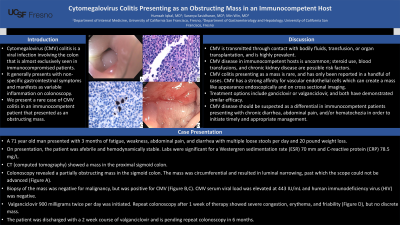
Case Description/Methods: A 71 year old man presented to the hospital with a 3 month history of fatigue, weakness, abdominal pain, and diarrhea with multiple loose stools per day and 20 pound weight loss. On presentation, the patient was afebrile and hemodynamically stable. Labs were significant for a Westergren sedimentation rate (ESR) 70 mm and C-reactive protein (CRP) 78.5 mg/L. Computed tomography (CT) showed a mass in the proximal sigmoid colon. Colonoscopy revealed a partially obstructing mass in the sigmoid colon. The mass was circumferential and resulted in luminal narrowing, past which the scope could not be advanced (Figure A). Biopsy of the mass was negative for malignancy, but was positive for CMV (Figure B,C). CMV serum viral load was elevated at 443 IU/mL and human immunodeficiency virus (HIV) was negative. Valganciclovir 900 milligrams twice per day was initiated. Repeat colonoscopy after 1 week of therapy showed severe congestion, erythema, and friability (Figure D), but no discrete mass. The patient was discharged with a 2 week course of valganciclovir and is pending repeat colonoscopy in 6 months.
Discussion: CMV is transmitted through contact with bodily fluids, transfusion, or organ transplantation, and is highly prevalent in the adult population. CMV disease in immunocompetent hosts is uncommon, and some studies identify steroid use, blood transfusions, and chronic kidney disease as possible risk factors. CMV colitis presenting as a mass is comparatively rare, and has only been reported in a handful of cases. This virus has a strong affinity for vascular endothelial cells which can create a mass like appearance endoscopically and on cross sectional imaging. All reported cases of CMV colitis presenting as a mass demonstrated resolution of the mass with appropriate antiviral therapy, as seen in our patient. Treatment options include ganciclovir or valganciclovir, and both have demonstrated similar efficacy. CMV disease should be suspected as a differential in immunocompetent patients presenting with chronic diarrhea, abdominal pain, and/or hematochezia in order to initiate timely and appropriate management.

Disclosures:
Humzah Iqbal, MD1, Saranya Sasidharan, MD2, Min Win, MD2. P1627 - Cytomegalovirus Colitis Presenting as an Obstructing Mass in an Immunocompetent Host, ACG 2023 Annual Scientific Meeting Abstracts. Vancouver, BC, Canada: American College of Gastroenterology.
1University of California San Francisco, Fresno, Fresno, CA; 2UCSF Fresno, Fresno, CA
Introduction: Cytomegalovirus (CMV) colitis is a viral infection involving the colon that is almost exclusively seen in immunocompromised patients. It generally presents with non-specific gastrointestinal symptoms and manifests as variable inflammation on colonoscopy. We present a rare case of CMV colitis in an immunocompetent patient that presented as an obstructing mass.
Case Description/Methods: A 71 year old man presented to the hospital with a 3 month history of fatigue, weakness, abdominal pain, and diarrhea with multiple loose stools per day and 20 pound weight loss. On presentation, the patient was afebrile and hemodynamically stable. Labs were significant for a Westergren sedimentation rate (ESR) 70 mm and C-reactive protein (CRP) 78.5 mg/L. Computed tomography (CT) showed a mass in the proximal sigmoid colon. Colonoscopy revealed a partially obstructing mass in the sigmoid colon. The mass was circumferential and resulted in luminal narrowing, past which the scope could not be advanced (Figure A). Biopsy of the mass was negative for malignancy, but was positive for CMV (Figure B,C). CMV serum viral load was elevated at 443 IU/mL and human immunodeficiency virus (HIV) was negative. Valganciclovir 900 milligrams twice per day was initiated. Repeat colonoscopy after 1 week of therapy showed severe congestion, erythema, and friability (Figure D), but no discrete mass. The patient was discharged with a 2 week course of valganciclovir and is pending repeat colonoscopy in 6 months.
Discussion: CMV is transmitted through contact with bodily fluids, transfusion, or organ transplantation, and is highly prevalent in the adult population. CMV disease in immunocompetent hosts is uncommon, and some studies identify steroid use, blood transfusions, and chronic kidney disease as possible risk factors. CMV colitis presenting as a mass is comparatively rare, and has only been reported in a handful of cases. This virus has a strong affinity for vascular endothelial cells which can create a mass like appearance endoscopically and on cross sectional imaging. All reported cases of CMV colitis presenting as a mass demonstrated resolution of the mass with appropriate antiviral therapy, as seen in our patient. Treatment options include ganciclovir or valganciclovir, and both have demonstrated similar efficacy. CMV disease should be suspected as a differential in immunocompetent patients presenting with chronic diarrhea, abdominal pain, and/or hematochezia in order to initiate timely and appropriate management.

Figure: Figure A: First colonoscopy showing circumferential narrowing in the sigmoid colon. Figure B: Hematoxylin & eosin (H&E) stained slide of sigmoid mass biopsy showing CMV. Figure C: Immunohistochemical staining of sigmoid mass showing positivity for CMV. Figure D: Repeat colonoscopy showing inflammation without a discrete mass in the sigmoid colon.
Disclosures:
Humzah Iqbal indicated no relevant financial relationships.
Saranya Sasidharan indicated no relevant financial relationships.
Min Win indicated no relevant financial relationships.
Humzah Iqbal, MD1, Saranya Sasidharan, MD2, Min Win, MD2. P1627 - Cytomegalovirus Colitis Presenting as an Obstructing Mass in an Immunocompetent Host, ACG 2023 Annual Scientific Meeting Abstracts. Vancouver, BC, Canada: American College of Gastroenterology.
