Monday Poster Session
Category: Colon
P1644 - Beware of the Great Mimicker - Gastrointestinal Basidiobolomycosis: A Rare and Underdiagnosed Fungal Infection
Monday, October 23, 2023
10:30 AM - 4:15 PM PT
Location: Exhibit Hall


Hamdan AlAwadhi, MD
Cleveland Clinic Abu Dhabi
Abu Dhabi, Abu Dhabi, United Arab Emirates
Presenting Author(s)
Hamdan AlAwadhi, MD1, Mahmoud El Kaissi, MD1, Ahmad Nusair, MD2
1Cleveland Clinic Abu Dhabi, Abu Dhabi, Abu Dhabi, United Arab Emirates; 2Marshall University Medical Hospital, Huntington, WV
Introduction: Gastrointestinal Basidiobolomycosis (GIB) is a rare and underreported fungal infection that is commonly seen in areas with hot warm arid climates. This disease can affect anywhere from stomach, small intestine, colon, and liver(1) We report the first case of GIB in an adult in the United Arab Emirates.
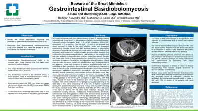
Case Description/Methods: A 31-year-old female with a history of type 1 diabetes mellitus was admitted to the hospital with a 1-day history of abdominal pain, nausea, and vomiting. Labs showed: WBC 11.5 x10*9/L, Hgb of 113 g/L, Plt of 573x10*9/L with eosinophilia 1.59 x10*9/L, CRP 76 mg/L. Liver enzymes, kidney function tests, and other electrolytes were all normal. CT scan of the abdomen and pelvis revealed a mass in the right paracolic gutter with associated inflammatory changes around the right ileocecal junction. A presumptive diagnosis of appendicular abscess was made, and the patient was started on conservative treatment of piperacillin/tazobactam. However, there was no improvement in her symptoms and so she underwent diagnostic laparoscopy. Intraoperative findings revealed a large appendicular tumor involving the cecum and ileocecal junction with multiple large regional lymph nodes with no peritoneal or liver metastasis and eventually, she had a right ileocecal resection. Histopathological identification of the mass revealed gangrenous perforating eosinophilic appendicitis with peri appendiceal abscess. Two weeks later she presented to the ED with worsening right-sided abdominal pain. CT scan of the abdomen and pelvis showed evidence of loculated thick-walled collection in the right paracolic gutter extending to the right side of the pelvis measuring 7.9x3.2 cm. Labs revealed a WBC 12.49 x10*9/L, Hgb of 81 g/L, Plt of 806 x10*9/L with eosinophilia of 2.86 x10*9/L.CRP 99.8 mg/L. Extensive workup done in light of the eosinophilia and thrombocytosis was nonrevealing. Additionally, she underwent extensive testing for infectious etiologies which were inconclusive. Eventually, the patient underwent aspiration of the fluid collection seen on the CT scan under radiological guidance. Surprisingly, her histopathology results revealed a Basidiobolus species growth in the fungal culture. She was started on a 6-month therapy of itraconazole with improvement in her clinical condition. Follow-up CT scans showed resolution of the fluid collection seen.
Discussion: This case highlights the importance of considering GIB as one of the differential diagnoses of mass-like lesions in the GI tract

Disclosures:
Hamdan AlAwadhi, MD1, Mahmoud El Kaissi, MD1, Ahmad Nusair, MD2. P1644 - Beware of the Great Mimicker - Gastrointestinal Basidiobolomycosis: A Rare and Underdiagnosed Fungal Infection, ACG 2023 Annual Scientific Meeting Abstracts. Vancouver, BC, Canada: American College of Gastroenterology.
1Cleveland Clinic Abu Dhabi, Abu Dhabi, Abu Dhabi, United Arab Emirates; 2Marshall University Medical Hospital, Huntington, WV
Introduction: Gastrointestinal Basidiobolomycosis (GIB) is a rare and underreported fungal infection that is commonly seen in areas with hot warm arid climates. This disease can affect anywhere from stomach, small intestine, colon, and liver(1) We report the first case of GIB in an adult in the United Arab Emirates.
Case Description/Methods: A 31-year-old female with a history of type 1 diabetes mellitus was admitted to the hospital with a 1-day history of abdominal pain, nausea, and vomiting. Labs showed: WBC 11.5 x10*9/L, Hgb of 113 g/L, Plt of 573x10*9/L with eosinophilia 1.59 x10*9/L, CRP 76 mg/L. Liver enzymes, kidney function tests, and other electrolytes were all normal. CT scan of the abdomen and pelvis revealed a mass in the right paracolic gutter with associated inflammatory changes around the right ileocecal junction. A presumptive diagnosis of appendicular abscess was made, and the patient was started on conservative treatment of piperacillin/tazobactam. However, there was no improvement in her symptoms and so she underwent diagnostic laparoscopy. Intraoperative findings revealed a large appendicular tumor involving the cecum and ileocecal junction with multiple large regional lymph nodes with no peritoneal or liver metastasis and eventually, she had a right ileocecal resection. Histopathological identification of the mass revealed gangrenous perforating eosinophilic appendicitis with peri appendiceal abscess. Two weeks later she presented to the ED with worsening right-sided abdominal pain. CT scan of the abdomen and pelvis showed evidence of loculated thick-walled collection in the right paracolic gutter extending to the right side of the pelvis measuring 7.9x3.2 cm. Labs revealed a WBC 12.49 x10*9/L, Hgb of 81 g/L, Plt of 806 x10*9/L with eosinophilia of 2.86 x10*9/L.CRP 99.8 mg/L. Extensive workup done in light of the eosinophilia and thrombocytosis was nonrevealing. Additionally, she underwent extensive testing for infectious etiologies which were inconclusive. Eventually, the patient underwent aspiration of the fluid collection seen on the CT scan under radiological guidance. Surprisingly, her histopathology results revealed a Basidiobolus species growth in the fungal culture. She was started on a 6-month therapy of itraconazole with improvement in her clinical condition. Follow-up CT scans showed resolution of the fluid collection seen.
Discussion: This case highlights the importance of considering GIB as one of the differential diagnoses of mass-like lesions in the GI tract

Figure: Image A: Axial CT scan of the abdomen and pelvis:
Star- Abnormal inflammatory/infectious changes seen around the right ileocecal junction with 7.9 x 3.2 cm collection in the right lower quadrant. The findings are worrisome for an abscess formation.
Image B: Coronal CT scan of the abdomen and pelvis:
7.9 x 3.2 cm collection in the right lower quadrant. The collection has a thick rim and central liquification process and necrosis.
Disclosures:
Hamdan AlAwadhi indicated no relevant financial relationships.
Mahmoud El Kaissi indicated no relevant financial relationships.
Ahmad Nusair indicated no relevant financial relationships.
Hamdan AlAwadhi, MD1, Mahmoud El Kaissi, MD1, Ahmad Nusair, MD2. P1644 - Beware of the Great Mimicker - Gastrointestinal Basidiobolomycosis: A Rare and Underdiagnosed Fungal Infection, ACG 2023 Annual Scientific Meeting Abstracts. Vancouver, BC, Canada: American College of Gastroenterology.
