Monday Poster Session
Category: Colon
P1649 - A Case of a Rectal Gastrointestinal Stromal Tumor (GIST): An Unexpected Cause of Lower GI Bleeding in an Immunocompromised Man
Monday, October 23, 2023
10:30 AM - 4:15 PM PT
Location: Exhibit Hall

Has Audio
- SW
Samantha Whitwell, MD
UTHSC
Memphis, TN
Presenting Author(s)
Samantha Whitwell, MD1, Kyle Kreitman, DO1, Claudio R.. Tombazzi, MD2
1UTHSC, Memphis, TN; 2Memphis Veterans Affairs Medical Center, Memphis, TN
Introduction: GISTs are rare, with only around 5,000 cases diagnosed annually in the United States. However, these tumors are the most common mesenchymal neoplasm in the GI tract. 5-15% of GISTS arise in the colon and rectum.
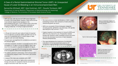
Case Description/Methods: A 49-year-old man with past medical history of acquired immune deficiency syndrome (AIDS, last CD4 count of 10 cells/mm3) presented with a 1-month history of daily epigastric pain and rectal bleeding. Physical exam was significant for condyloma acuminata in the perianal region. Laboratory evaluation revealed a normocytic anemia with a hemoglobin of 11.9 g/dL (normal 12-16g/dL), which was down from the patient’s baseline hemoglobin of 14g/dL. A CT abdomen/pelvis displayed acute nonspecific colitis of the rectosigmoid colon. The patient underwent colonoscopy which showed a 6 cm non-obstructive, polypoid mass in the rectum with central ulceration. A biopsy of the mass was consistent with a GIST. The patient was restarted on highly active antiretroviral therapy with plans for outpatient Imatinib and potential surgery later down the road.
Discussion: Most GISTs are diagnosed incidentally on imaging, endoscopy, or during a surgical procedure. GISTs are comprised of three histological categories including spindle, epithelioid, and mixed type, with spindle type being the most common (70%). Surgical resection is the treatment of choice for GISTs and offers the only chance for cure. Preoperative medical therapy with Imatinib is often indicated to shrink the tumor and can also decrease recurrence or treat metastatic disease posteroperatively. This case highlights the importance of pursing endoscopy in immunocompromised patients with nonspecific imaging findings to secure the diagnosis.
References
1. Mehren, Margaret von. “Gastrointestinal Stromal Tumors .” Sleisenger and Fordtran's Gastrointestinal and Liver Disease, Elsevier
2. Morgan, Jeffery, and Chandrajit Raut. “Clinical Presentation, Diagnosis, and Prognosis of Gastrointestinal Stromal Tumors.” UpToDate, Wolter Kluwer.
3. Morgan, Jeffery, and Chandrajit Raut. “Local Treatment for Gastrointestinal Stromal Tumors, Leiomyomas, and Leiomyosarcomas of the Gastrointestinal Tract.” UpToDate, Wolters Kluwer
4. von Mehren, Margaret, and John Kane. “Treatment by Cancer Type- Gastrointestinal Stromal Tumors (GISTs).” NCCN, NCCN,

Disclosures:
Samantha Whitwell, MD1, Kyle Kreitman, DO1, Claudio R.. Tombazzi, MD2. P1649 - A Case of a Rectal Gastrointestinal Stromal Tumor (GIST): An Unexpected Cause of Lower GI Bleeding in an Immunocompromised Man, ACG 2023 Annual Scientific Meeting Abstracts. Vancouver, BC, Canada: American College of Gastroenterology.
1UTHSC, Memphis, TN; 2Memphis Veterans Affairs Medical Center, Memphis, TN
Introduction: GISTs are rare, with only around 5,000 cases diagnosed annually in the United States. However, these tumors are the most common mesenchymal neoplasm in the GI tract. 5-15% of GISTS arise in the colon and rectum.
Case Description/Methods: A 49-year-old man with past medical history of acquired immune deficiency syndrome (AIDS, last CD4 count of 10 cells/mm3) presented with a 1-month history of daily epigastric pain and rectal bleeding. Physical exam was significant for condyloma acuminata in the perianal region. Laboratory evaluation revealed a normocytic anemia with a hemoglobin of 11.9 g/dL (normal 12-16g/dL), which was down from the patient’s baseline hemoglobin of 14g/dL. A CT abdomen/pelvis displayed acute nonspecific colitis of the rectosigmoid colon. The patient underwent colonoscopy which showed a 6 cm non-obstructive, polypoid mass in the rectum with central ulceration. A biopsy of the mass was consistent with a GIST. The patient was restarted on highly active antiretroviral therapy with plans for outpatient Imatinib and potential surgery later down the road.
Discussion: Most GISTs are diagnosed incidentally on imaging, endoscopy, or during a surgical procedure. GISTs are comprised of three histological categories including spindle, epithelioid, and mixed type, with spindle type being the most common (70%). Surgical resection is the treatment of choice for GISTs and offers the only chance for cure. Preoperative medical therapy with Imatinib is often indicated to shrink the tumor and can also decrease recurrence or treat metastatic disease posteroperatively. This case highlights the importance of pursing endoscopy in immunocompromised patients with nonspecific imaging findings to secure the diagnosis.
References
1. Mehren, Margaret von. “Gastrointestinal Stromal Tumors .” Sleisenger and Fordtran's Gastrointestinal and Liver Disease, Elsevier
2. Morgan, Jeffery, and Chandrajit Raut. “Clinical Presentation, Diagnosis, and Prognosis of Gastrointestinal Stromal Tumors.” UpToDate, Wolter Kluwer.
3. Morgan, Jeffery, and Chandrajit Raut. “Local Treatment for Gastrointestinal Stromal Tumors, Leiomyomas, and Leiomyosarcomas of the Gastrointestinal Tract.” UpToDate, Wolters Kluwer
4. von Mehren, Margaret, and John Kane. “Treatment by Cancer Type- Gastrointestinal Stromal Tumors (GISTs).” NCCN, NCCN,

Figure: Top Left: CT with contrast showing nonspecific colitis pattern
Top RIght: Endoscopic imaging with non-obstructive, polypoid mass with central ulceration
Bottom Left: (Narrow band imaging): Endoscopic images with non-obstructive, polypoid mass with central ulceration.
Bottom Right: Histological Findings of GIST with mixed phenotype, showing both spindle and epithelioid cell types
Disclosures:
Samantha Whitwell indicated no relevant financial relationships.
Kyle Kreitman indicated no relevant financial relationships.
Claudio Tombazzi indicated no relevant financial relationships.
Samantha Whitwell, MD1, Kyle Kreitman, DO1, Claudio R.. Tombazzi, MD2. P1649 - A Case of a Rectal Gastrointestinal Stromal Tumor (GIST): An Unexpected Cause of Lower GI Bleeding in an Immunocompromised Man, ACG 2023 Annual Scientific Meeting Abstracts. Vancouver, BC, Canada: American College of Gastroenterology.
