Monday Poster Session
Category: Colon
P1657 - Melanoma in the Shadows: A Case Report of Anorectal Malignant Melanoma
Monday, October 23, 2023
10:30 AM - 4:15 PM PT
Location: Exhibit Hall

Has Audio

Paul Kozak, DO
Riverside Medical Center
Kankakee, IL
Presenting Author(s)
Paul Kozak, DO1, Alexander Satinsky, MS2, Daniel Errampalli, MD3
1Riverside Medical Center, Kankakee, IL; 2Chicago College of Osteopathic Medicine, Midwestern University, Downers Grove, IL; 3Digestive Disease Consultants, Bourbonnais, IL
Introduction: While classically a dermatologic malignancy, melanoma is cutaneous only 91.2% of the time. It seldom occurs on mucosal sites (1.3%), and more rarely in the anorectum (0.3%) where early detection is crucial for prognosis.1,2
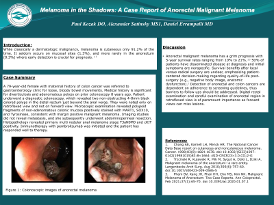
Case Description/Methods: A 79-year-old female with maternal history of colon cancer was referred to gastroenterology clinic for loose, bloody bowel movements. Medical history is significant for diverticulosis and adenomatous polyps on prior colonoscopy 8 years ago. Patient underwent a diagnostic colonoscopy, which revealed two non-obstructing 4-8mm black colored polyps in the distal rectum just beyond the anal verge. They were originally identified in the retroflexed view. Microscopic examination revealed polypoid fragments of non-adenomatous colonic mucosa positively stained with MART1, SOX10, and Tyrosinase, consistent with margin positive malignant melanoma. Imaging studies did not reveal metastasis, and she subsequently underwent abdominoperineal resection. Histopathology revealed primary multinodular anal melanoma stage T3aN0M0 and cKIT positivity. Immunotherapy with pembrolizumab was initiated and the patient has responded well to therapy.
Discussion: Anorectal malignant melanoma has a grim prognosis with 5-year survival rates ranging from 10% to 21%.2,3 50% of patients have disseminated disease at diagnosis and initial symptoms are nonspecific. Survival benefits after local versus radical surgery are unclear, emphasizing patient-centered decision-making regarding quality-of-life post-surgery (e.g., negative body image, anatomic dysfunction).2 Detection of anorectal and colon cancers are dependent on adherence to screening guidelines, thus barriers to follow ups should be addressed. Digital rectal examination and careful examination of anorectal region in retroflexed view is of paramount importance as forward views can miss lesions.
References:
1.
Chang AE, Karnell LH, Menck HR.
Base report on cutaneous and noncutaneous melanoma. Cancer.
1998;83(8):1664-1678.
2.
Trzcinski R, Kujawski R, Mik M, Sygut A, Dziki L, Dziki A.
Malignant melanoma of the anorectum--a rare entity.
Langenbecks Arch Surg. Aug 2010;395(6):757-60.
3.
Pham BV, Kang JH, Phan HH, Cho MS, Kim NK. Malignant
Melanoma of Anorectum: Two Case Reports. Ann Coloproctol.
Feb 2021;37(1):65-70.

Disclosures:
Paul Kozak, DO1, Alexander Satinsky, MS2, Daniel Errampalli, MD3. P1657 - Melanoma in the Shadows: A Case Report of Anorectal Malignant Melanoma, ACG 2023 Annual Scientific Meeting Abstracts. Vancouver, BC, Canada: American College of Gastroenterology.
1Riverside Medical Center, Kankakee, IL; 2Chicago College of Osteopathic Medicine, Midwestern University, Downers Grove, IL; 3Digestive Disease Consultants, Bourbonnais, IL
Introduction: While classically a dermatologic malignancy, melanoma is cutaneous only 91.2% of the time. It seldom occurs on mucosal sites (1.3%), and more rarely in the anorectum (0.3%) where early detection is crucial for prognosis.1,2
Case Description/Methods: A 79-year-old female with maternal history of colon cancer was referred to gastroenterology clinic for loose, bloody bowel movements. Medical history is significant for diverticulosis and adenomatous polyps on prior colonoscopy 8 years ago. Patient underwent a diagnostic colonoscopy, which revealed two non-obstructing 4-8mm black colored polyps in the distal rectum just beyond the anal verge. They were originally identified in the retroflexed view. Microscopic examination revealed polypoid fragments of non-adenomatous colonic mucosa positively stained with MART1, SOX10, and Tyrosinase, consistent with margin positive malignant melanoma. Imaging studies did not reveal metastasis, and she subsequently underwent abdominoperineal resection. Histopathology revealed primary multinodular anal melanoma stage T3aN0M0 and cKIT positivity. Immunotherapy with pembrolizumab was initiated and the patient has responded well to therapy.
Discussion: Anorectal malignant melanoma has a grim prognosis with 5-year survival rates ranging from 10% to 21%.2,3 50% of patients have disseminated disease at diagnosis and initial symptoms are nonspecific. Survival benefits after local versus radical surgery are unclear, emphasizing patient-centered decision-making regarding quality-of-life post-surgery (e.g., negative body image, anatomic dysfunction).2 Detection of anorectal and colon cancers are dependent on adherence to screening guidelines, thus barriers to follow ups should be addressed. Digital rectal examination and careful examination of anorectal region in retroflexed view is of paramount importance as forward views can miss lesions.
References:
1.
Chang AE, Karnell LH, Menck HR.
Base report on cutaneous and noncutaneous melanoma. Cancer.
1998;83(8):1664-1678.
2.
Trzcinski R, Kujawski R, Mik M, Sygut A, Dziki L, Dziki A.
Malignant melanoma of the anorectum--a rare entity.
Langenbecks Arch Surg. Aug 2010;395(6):757-60.
3.
Pham BV, Kang JH, Phan HH, Cho MS, Kim NK. Malignant
Melanoma of Anorectum: Two Case Reports. Ann Coloproctol.
Feb 2021;37(1):65-70.

Figure: Figure 1: Colonoscopic image of anorectal melanoma
Disclosures:
Paul Kozak indicated no relevant financial relationships.
Alexander Satinsky indicated no relevant financial relationships.
Daniel Errampalli indicated no relevant financial relationships.
Paul Kozak, DO1, Alexander Satinsky, MS2, Daniel Errampalli, MD3. P1657 - Melanoma in the Shadows: A Case Report of Anorectal Malignant Melanoma, ACG 2023 Annual Scientific Meeting Abstracts. Vancouver, BC, Canada: American College of Gastroenterology.
