Monday Poster Session
Category: Colon
P1690 - It is Not Just Fibrous Adhesions
Monday, October 23, 2023
10:30 AM - 4:15 PM PT
Location: Exhibit Hall

Has Audio

Raj Jessica Thomas, DO
Cleveland Clinic
Cleveland, OH
Presenting Author(s)
Raj Jessica Thomas, DO1, Stephanie Washburn, BS2, Vishal M. Patel, MD3, Douglas J.. Grider, MD4
1Cleveland Clinic Akron General, Akron, OH; 2Edward Via College of Osteopathic Medicine, Blacksburg, VA; 3Carilion Roanoke Memorial Hospital, Roanoke, VA; 4Virginia Tech Carilion School of Medicine, Roanoke, VA
Introduction: Inflammatory myofibroblastic tumor (IMT) is rare mesenchymal tumor composed of myofibroblastic spindle cells admixed with inflammatory cells. The etiology of IMT remain uncertain. They generally occur in the lung, mesentery, and omentum. There is a high propensity of local recurrence and only metastasize in rare cases. Anaplastic lymphoma kinase (ALK) rearrangements and/or ALK1 and p80 immunoreactivity are reported in more than half the cases. Surgical resection remains the treatment of choice and when ALK positive tumors are found ALK inhibitors are preferred.
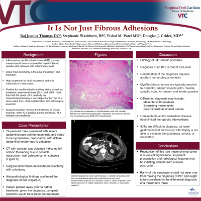
Case Description/Methods: This report describes a 72-year-old male who presented with severe abdominal pain and hematochezia, CT imaging revealed pancolitis suspicious for ischemic versus infectious colitis. He was treated with medical management in the setting of ischemic colitis. A week into the hospital course, patient had reoccurrence of abdominal pain. CT with contrast was obtained indicative of left colonic thickening due to possible obstruction, wall dehiscence, or ischemia(Figure 1B.) Given this surgical intervention was pursued, findings were notable for: full thickness ischemia of distal transverse colon to proximal sigmoid colon with extensive gross adhesions noted at the splenic flexure and throughout the small bowel. Histopathology of the colon was evident for transmural necrosis, ischemic changes, and fibrous adhesions. Proliferation of larger spindle myofibroblastic cells intermixed with lymphocytes, histocytes, and plasma cells extending from the subserosa into the muscularis propria were noted (Figure 1A); spindle cells were smooth muscle actin (SMA) and keratin positive. Histopathology of the small bowel was evident of spindle cell proliferation into the serosa and subserosa and was SMA, keratin, and focally desmin positive. The substantial overtaking of the IMT was compressive in nature to the mesentery resulting in bowel ischemia. ALK immunoreactivity was noted to be negative.
Discussion: In essence, recognition of this rare mesenchymal tumor is of significance, as clinical manifestation and radiological features may be indistinguishable from a bowel obstruction. Differential diagnosis may include mesenteric fibromatosis, sclerosing mesenteritis, and gastrointestinal stromal tumor. Rarity of this neoplasm should not deter one from making the diagnosis of IMT and ought to be considered in the differential diagnosis for a mesenteric mass.

Disclosures:
Raj Jessica Thomas, DO1, Stephanie Washburn, BS2, Vishal M. Patel, MD3, Douglas J.. Grider, MD4. P1690 - It is Not Just Fibrous Adhesions, ACG 2023 Annual Scientific Meeting Abstracts. Vancouver, BC, Canada: American College of Gastroenterology.
1Cleveland Clinic Akron General, Akron, OH; 2Edward Via College of Osteopathic Medicine, Blacksburg, VA; 3Carilion Roanoke Memorial Hospital, Roanoke, VA; 4Virginia Tech Carilion School of Medicine, Roanoke, VA
Introduction: Inflammatory myofibroblastic tumor (IMT) is rare mesenchymal tumor composed of myofibroblastic spindle cells admixed with inflammatory cells. The etiology of IMT remain uncertain. They generally occur in the lung, mesentery, and omentum. There is a high propensity of local recurrence and only metastasize in rare cases. Anaplastic lymphoma kinase (ALK) rearrangements and/or ALK1 and p80 immunoreactivity are reported in more than half the cases. Surgical resection remains the treatment of choice and when ALK positive tumors are found ALK inhibitors are preferred.
Case Description/Methods: This report describes a 72-year-old male who presented with severe abdominal pain and hematochezia, CT imaging revealed pancolitis suspicious for ischemic versus infectious colitis. He was treated with medical management in the setting of ischemic colitis. A week into the hospital course, patient had reoccurrence of abdominal pain. CT with contrast was obtained indicative of left colonic thickening due to possible obstruction, wall dehiscence, or ischemia(Figure 1B.) Given this surgical intervention was pursued, findings were notable for: full thickness ischemia of distal transverse colon to proximal sigmoid colon with extensive gross adhesions noted at the splenic flexure and throughout the small bowel. Histopathology of the colon was evident for transmural necrosis, ischemic changes, and fibrous adhesions. Proliferation of larger spindle myofibroblastic cells intermixed with lymphocytes, histocytes, and plasma cells extending from the subserosa into the muscularis propria were noted (Figure 1A); spindle cells were smooth muscle actin (SMA) and keratin positive. Histopathology of the small bowel was evident of spindle cell proliferation into the serosa and subserosa and was SMA, keratin, and focally desmin positive. The substantial overtaking of the IMT was compressive in nature to the mesentery resulting in bowel ischemia. ALK immunoreactivity was noted to be negative.
Discussion: In essence, recognition of this rare mesenchymal tumor is of significance, as clinical manifestation and radiological features may be indistinguishable from a bowel obstruction. Differential diagnosis may include mesenteric fibromatosis, sclerosing mesenteritis, and gastrointestinal stromal tumor. Rarity of this neoplasm should not deter one from making the diagnosis of IMT and ought to be considered in the differential diagnosis for a mesenteric mass.

Figure: Figure 1: (A) Spindle cell proliferation of myofibroblastic cells with variable fibrosis and intermixed lymphocytes, histocytes, and plasma cells into the muscularis propria [H&E 20X magnification]. (B) Normal proximal colon wall thickness (1.) Distal transverse colon and proximal descending colon wall thickening and hypo enhancement suspicious for ischemic colitis (2.) Point of partial colon obstruction and no visible obstructive colon, omental, or mesenteric mass (3.)
Disclosures:
Raj Jessica Thomas indicated no relevant financial relationships.
Stephanie Washburn indicated no relevant financial relationships.
Vishal Patel indicated no relevant financial relationships.
Douglas Grider indicated no relevant financial relationships.
Raj Jessica Thomas, DO1, Stephanie Washburn, BS2, Vishal M. Patel, MD3, Douglas J.. Grider, MD4. P1690 - It is Not Just Fibrous Adhesions, ACG 2023 Annual Scientific Meeting Abstracts. Vancouver, BC, Canada: American College of Gastroenterology.
