Monday Poster Session
Category: Colon
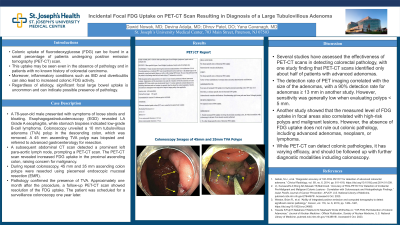
P1743 - Incidental Focal FDG Uptake on PET-CT Scan Resulting in Diagnosis of a Large Tubulovillous Adenoma
Monday, October 23, 2023
10:30 AM - 4:15 PM PT
Location: Exhibit Hall

Has Audio
- DN
Dawid Nowak, MD
St. Joseph's University Medical Center
Paterson, NJ
Presenting Author(s)
Dawid Nowak, MD1, Devina Adalja, MD1, Dhruv Patel, DO2, Yana Cavanagh, MD2
1St. Joseph's University Medical Center, Paterson, NJ; 2St. Joseph's University Medical Center, Paterson, NJ
Introduction: Colonic uptake of fluorodeoxyglucose (FDG) can be found in a small percentage of patients undergoing positron emission tomography (PET-CT) scan. This uptake may be seen even in the absence of pathology and in patients with no known history of colorectal carcinoma. Moreover, inflammatory conditions such as IBD and diverticulitis can also lead to increased colonic FDG activity. Regardless of etiology, significant focal large bowel uptake is uncommon and can indicate possible presence of pathology.
Case Description/Methods: A 78-year-old male presented with symptoms of loose stools and bloating. Esophagogastroduodenoscopy (EGD) revealed LA Grade A esophagitis, while stomach biopsies indicated low-grade B-cell lymphoma. Colonoscopy unveiled a 10 mm tubulovillous adenoma (TVA) polyp in the descending colon, which was removed. A 45 mm ascending TVA polyp was biopsied and referred to advanced gastroenterology for resection. A subsequent abdominal CT scan detected a prominent left para-aortic lymph node, prompting a PET-CT scan. The PET-CT scan revealed increased FDG uptake in the proximal ascending colon, raising concern for malignancy. During repeat colonoscopy, 45 mm and 35 mm ascending colon polyps were resected using piecemeal endoscopic mucosal resection (EMR). Pathology confirmed the presence of TVA. Approximately one month after the procedure, a follow-up PET-CT scan showed resolution of the FDG uptake. The patient was scheduled for a surveillance colonoscopy one year later.
Discussion: Several studies have assessed the effectiveness of PET-CT scans in detecting colorectal pathology, with one study finding that PET-CT scans identified only about half of patients with advanced adenomas. The detection rate of PET imaging correlated with the size of the adenomas, with a 90% detection rate for adenomas ≥ 13 mm in another study. However, sensitivity was generally low when evaluating polyps < 5 mm. Another study showed that the measured level of FDG uptake in focal areas also correlated with high-risk polyps and malignant lesions. However, the absence of FDG uptake does not rule out colonic pathology, including advanced adenomas, neoplasm, or lymphoma. While PET-CT can detect colonic pathologies, it has varying efficacy, and should be followed up with further diagnostic modalities including colonoscopy.
Disclosures:
Dawid Nowak, MD1, Devina Adalja, MD1, Dhruv Patel, DO2, Yana Cavanagh, MD2. P1743 - Incidental Focal FDG Uptake on PET-CT Scan Resulting in Diagnosis of a Large Tubulovillous Adenoma, ACG 2023 Annual Scientific Meeting Abstracts. Vancouver, BC, Canada: American College of Gastroenterology.
1St. Joseph's University Medical Center, Paterson, NJ; 2St. Joseph's University Medical Center, Paterson, NJ
Introduction: Colonic uptake of fluorodeoxyglucose (FDG) can be found in a small percentage of patients undergoing positron emission tomography (PET-CT) scan. This uptake may be seen even in the absence of pathology and in patients with no known history of colorectal carcinoma. Moreover, inflammatory conditions such as IBD and diverticulitis can also lead to increased colonic FDG activity. Regardless of etiology, significant focal large bowel uptake is uncommon and can indicate possible presence of pathology.
Case Description/Methods: A 78-year-old male presented with symptoms of loose stools and bloating. Esophagogastroduodenoscopy (EGD) revealed LA Grade A esophagitis, while stomach biopsies indicated low-grade B-cell lymphoma. Colonoscopy unveiled a 10 mm tubulovillous adenoma (TVA) polyp in the descending colon, which was removed. A 45 mm ascending TVA polyp was biopsied and referred to advanced gastroenterology for resection. A subsequent abdominal CT scan detected a prominent left para-aortic lymph node, prompting a PET-CT scan. The PET-CT scan revealed increased FDG uptake in the proximal ascending colon, raising concern for malignancy. During repeat colonoscopy, 45 mm and 35 mm ascending colon polyps were resected using piecemeal endoscopic mucosal resection (EMR). Pathology confirmed the presence of TVA. Approximately one month after the procedure, a follow-up PET-CT scan showed resolution of the FDG uptake. The patient was scheduled for a surveillance colonoscopy one year later.
Discussion: Several studies have assessed the effectiveness of PET-CT scans in detecting colorectal pathology, with one study finding that PET-CT scans identified only about half of patients with advanced adenomas. The detection rate of PET imaging correlated with the size of the adenomas, with a 90% detection rate for adenomas ≥ 13 mm in another study. However, sensitivity was generally low when evaluating polyps < 5 mm. Another study showed that the measured level of FDG uptake in focal areas also correlated with high-risk polyps and malignant lesions. However, the absence of FDG uptake does not rule out colonic pathology, including advanced adenomas, neoplasm, or lymphoma. While PET-CT can detect colonic pathologies, it has varying efficacy, and should be followed up with further diagnostic modalities including colonoscopy.
Disclosures:
Dawid Nowak indicated no relevant financial relationships.
Devina Adalja indicated no relevant financial relationships.
Dhruv Patel indicated no relevant financial relationships.
Yana Cavanagh indicated no relevant financial relationships.
Dawid Nowak, MD1, Devina Adalja, MD1, Dhruv Patel, DO2, Yana Cavanagh, MD2. P1743 - Incidental Focal FDG Uptake on PET-CT Scan Resulting in Diagnosis of a Large Tubulovillous Adenoma, ACG 2023 Annual Scientific Meeting Abstracts. Vancouver, BC, Canada: American College of Gastroenterology.
