Monday Poster Session
Category: Endoscopy Video Forum
P1796 - Novel Use of Self-Assembling Peptide Hemostatic Gel for Hemostasis of Pancreas Intracystic Bleeding

- SU
Shifa Umar, MD
University of Chicago
Chicago, Illinois
Presenting Author(s)
1University of Chicago, Chicago, IL; 2Center for Endoscopic Research and Therapeutics (CERT), University of Chicago, Chicago, IL
Introduction:
Endoscopic ultrasound (EUS) guided fine needle aspiration (FNA) of pancreas cyst is a commonly performed procedure. Adverse events for EUS guided FNA include luminal and intralesional bleeding (less than 1%), however there is a paucity of endoscopically available tools and techniques for management of intralesional bleeding during EUS FNA. We describe a novel technique utilizing self-assembling peptide hemostatic gel for management of intracystic bleeding in a pancreas cyst following EUS-guided FNA.
Case Description/Methods:
A 67-year-old female with end stage renal disease on hemodialysis being evaluated for renal transplant was referred for an incidental finding of a 2.1 x 2.6 cm uncinate pancreas cyst associated with pancreas duct dilation up to 4 mm noted on MRCP.
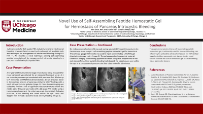
EUS demonstrated an anechoic lesion suggestive of a cyst measuring 2.6 cm in the uncinate process of the pancreas with a small intracystic mucin globule (Image 1). The cyst was noted to be in communication with the pancreatic duct. There were no septations, debris, or associated mass, and the outer wall of the lesion was thin. Before the FNA, color doppler imaging was utilized to confirm a lack of significant vascular structures within the needle path. One pass was made with a 22-gauge FNA needle using a transduodenal approach. No stylet was used. Immediately following puncture, active bleeding was noted into the cyst cavity and 0.2mL of bloody cystic fluid was safely aspirated (Image 2). Fluid sample was sent for cytology. To stop the bleeding,1 ml of self-assembling peptide hemostatic gel was injected into the cystic cavity using a 22-gauge needle (Image 3). The cystic lesion was observed endosonographically and all active bleeding appeared to stop. No bleeding or blood seen within the cyst or at the duodenal puncture site after observation for 15 minutes.
Cytology demonstrated bland mucinous glandular cells in a background of cyst content with no evidence of dysplasia or malignancy. Likely diagnosis remains side branch IPMN. The patient reported doing well with no post-procedural complications noted as of her 3 month outpatient follow-up with nephrology.
Discussion:
This case demonstrates that a self-assembling peptide hemostatic gel, traditionally used for mucosal bleeding, can safely and effectively be utilized to achieve hemostasis for FNA related bleeding inside a pancreatic cyst. Larger studies could further validate the use of self-assembling peptide hemostatic gel to treat bleeding inside pancreatic cystic lesions.

Image 2. Intracystic bleeding following FNA needle puncture
Image 3. Self-assembling peptide hemostatic gel was injected into the cystic cavity using a 22 gauge FNA needle
Disclosures:
Shifa Umar, MD1, Grace E.. Kim, MD1, Uzma D. Siddiqui, MD2. P1796 - Novel Use of Self-Assembling Peptide Hemostatic Gel for Hemostasis of Pancreas Intracystic Bleeding, ACG 2023 Annual Scientific Meeting Abstracts. Vancouver, BC, Canada: American College of Gastroenterology.
