Sunday Poster Session
Category: Biliary/Pancreas
P0101 - A Rare Ca(u)se of Liver Abscess
Sunday, October 22, 2023
3:30 PM - 7:00 PM PT
Location: Exhibit Hall

Has Audio
- SP
Sailaja Pisipati, MBBS
Mayo Clinic Arizona
Rochester, MN
Presenting Author(s)
Sailaja Pisipati, MBBS1, Michelle Anderson, MD2, Rahul Pannala, MD2, Douglas O. Faigel, MD2
1Mayo Clinic Arizona, Scottsdale, AZ; 2Mayo Clinic, Scottsdale, AZ
Introduction: Choledochal cysts (CDCs) are rare congenital dilatations of various segments of the intra-hepatic and/or extrahepatic biliary tract, affecting about 1 in 100000 live births in the West. The majority of CDCs are identified within the first decade of life. When left undiagnosed into adulthood, they are associated with increased risk of malignancy.
Case Description/Methods: 70-year-old Native American female presented with 2-weeks of abdominal pain, associated with nausea and anorexia. Labs suggested elevated alkaline phosphatase 203 and lipase > 4000. Noncontrast CT abdomen demonstrated peripancreatic stranding in keeping with mild pancreatitis, gallbladder wall thickening with possible perforation resulting in a hepatic abscess 44 x 18 x 29 mm, and dilated common bile duct (CBD) 21 mm. Following intial resuscitation with fluids and antibiotics, MRCP was obtained. This showed features of acute interstitial pancreatitis, mildly enlarged peripancreatic lymph nodes, distended gall bladder with diffuse wall thickening and enhancement, focal discontinuity indicating perforation and resultant liver abscess. Proximal CBD was dilated to 21 mm, with abrupt, smooth narrowing of 2.5 cm of distal CBD. Endoscopic retrograde cholangiopancreatography (ERCP) and endoscopic ultrasound (EUS) were deferred. Percutaneous drainage of gall bladder was considered but patient underwent cholecystectomy. Pathology revealed poorly differentiated, diffusely invasive adenocarcinoma on background of extensive high-grade dysplasia, positive cystic duct margin and lymphovascular invasion. Subsequent EUS revealed dilated CBD 20 mm, diffuse echogenicity of pancreas, malignant-appearing lymph nodes. Cholangiogram demonstrated fusiform dilation of CBD in keeping with type I CDC and a 20 mm common pancreatico-biliary channel with anomalous pancreatobiliary junction (APBJ). Patient unfortunately developed metastatic disease with malignant ascites and elected home hospice.
Discussion: CDCs are thought to arise due to APBJ with a long common channel, leading to reflux of pancreatic secretions into the biliary tract with resultant injury and cyst formation. Complications of CDCs include biliary stones, recurrent cholangitis, pancreatitis, biliary and hepatic fibrosis, and malignancy. Risk of malignancy increases with age, varies from 2.5% to 21%, and is highest in type 1 CDC. Early diagnosis and surgical excision with biliary diversion is recommended for management of type 1 CDC.

Disclosures:
Sailaja Pisipati, MBBS1, Michelle Anderson, MD2, Rahul Pannala, MD2, Douglas O. Faigel, MD2. P0101 - A Rare Ca(u)se of Liver Abscess, ACG 2023 Annual Scientific Meeting Abstracts. Vancouver, BC, Canada: American College of Gastroenterology.
1Mayo Clinic Arizona, Scottsdale, AZ; 2Mayo Clinic, Scottsdale, AZ
Introduction: Choledochal cysts (CDCs) are rare congenital dilatations of various segments of the intra-hepatic and/or extrahepatic biliary tract, affecting about 1 in 100000 live births in the West. The majority of CDCs are identified within the first decade of life. When left undiagnosed into adulthood, they are associated with increased risk of malignancy.
Case Description/Methods: 70-year-old Native American female presented with 2-weeks of abdominal pain, associated with nausea and anorexia. Labs suggested elevated alkaline phosphatase 203 and lipase > 4000. Noncontrast CT abdomen demonstrated peripancreatic stranding in keeping with mild pancreatitis, gallbladder wall thickening with possible perforation resulting in a hepatic abscess 44 x 18 x 29 mm, and dilated common bile duct (CBD) 21 mm. Following intial resuscitation with fluids and antibiotics, MRCP was obtained. This showed features of acute interstitial pancreatitis, mildly enlarged peripancreatic lymph nodes, distended gall bladder with diffuse wall thickening and enhancement, focal discontinuity indicating perforation and resultant liver abscess. Proximal CBD was dilated to 21 mm, with abrupt, smooth narrowing of 2.5 cm of distal CBD. Endoscopic retrograde cholangiopancreatography (ERCP) and endoscopic ultrasound (EUS) were deferred. Percutaneous drainage of gall bladder was considered but patient underwent cholecystectomy. Pathology revealed poorly differentiated, diffusely invasive adenocarcinoma on background of extensive high-grade dysplasia, positive cystic duct margin and lymphovascular invasion. Subsequent EUS revealed dilated CBD 20 mm, diffuse echogenicity of pancreas, malignant-appearing lymph nodes. Cholangiogram demonstrated fusiform dilation of CBD in keeping with type I CDC and a 20 mm common pancreatico-biliary channel with anomalous pancreatobiliary junction (APBJ). Patient unfortunately developed metastatic disease with malignant ascites and elected home hospice.
Discussion: CDCs are thought to arise due to APBJ with a long common channel, leading to reflux of pancreatic secretions into the biliary tract with resultant injury and cyst formation. Complications of CDCs include biliary stones, recurrent cholangitis, pancreatitis, biliary and hepatic fibrosis, and malignancy. Risk of malignancy increases with age, varies from 2.5% to 21%, and is highest in type 1 CDC. Early diagnosis and surgical excision with biliary diversion is recommended for management of type 1 CDC.

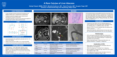
Figure: a) MRI with diffusely thickened and enhancing gall bladder wall with focal discontinuity
b) 1.5 x 4.5 cm collection with peripheral enhancement
c) Retrospective review of MRI images demonstrating fusiform dilation of bile duct, in keeping with Type 1 Choledochal Cyst (green arrow)
d) Retrospective review of the MRCP demonstrating type 1 Choledochal Cyst (green arrow), APBJ (red arrow) and pancreatic duct (yellow arrow)
e) Histopathology showing poorly differentiated, diffusely invasive adenocarcinoma of gall bladder with high grade dysplasia
f) Cholangiogram demonstrating Type 1 Choledochal Cyst (green arrow) and long common channel (yellow arrow)
Disclosures:
Sailaja Pisipati indicated no relevant financial relationships.
Michelle Anderson indicated no relevant financial relationships.
Rahul Pannala: Aimmune Therapeutics, a Nestlé Health Science company – Consultant. blue star genomics – Advisor or Review Panel Member. HCL technologies – Consultant.
Douglas Faigel indicated no relevant financial relationships.
Sailaja Pisipati, MBBS1, Michelle Anderson, MD2, Rahul Pannala, MD2, Douglas O. Faigel, MD2. P0101 - A Rare Ca(u)se of Liver Abscess, ACG 2023 Annual Scientific Meeting Abstracts. Vancouver, BC, Canada: American College of Gastroenterology.
