Sunday Poster Session
Category: Biliary/Pancreas
P0105 - Mucinous Adenocarcinoma of the Gallbladder with Signet Ring Cells: A Rare Histopathological Phenomenon
Sunday, October 22, 2023
3:30 PM - 7:00 PM PT
Location: Exhibit Hall

Has Audio

Nehal Patel, MD
Rosalind Franklin University of Medicine and Science
Chicago, IL
Presenting Author(s)
Award: Presidential Poster Award
Swetha Paduri, MD1, Nehal Patel, MD1, Jessica Eisold, MD2, Haoliang Xu, MD3, Liam Nolan, MD3, Aditya Gupta, MD3
1Rosalind Franklin University of Medicine and Science, Chicago, IL; 2American University of the Caribbean, Chicago, IL; 3Mount Sinai Hospital, Chicago, IL
Introduction: Gallbladder carcinomas are relatively uncommon. The majority of gallbladder tumors are classified as adenocarcinomas. Mucinous carcinoma (MC) of the gallbladder is a rare histopathological variant and signet-ring cells are seldom seen in them. This is a unique case that was diagnosed incidentally on post-operative specimen evaluation.
Case Description/Methods: A 65-year-old male with a history of hypertension, opioid use disorder on methadone presented with a two-day history of abdominal pain accompanied with nausea and bilious vomiting. Laboratory results were significant for leukocytosis, hyperbilirubinemia, and elevated liver associated enzymes. Imaging was concerning for biliary obstruction. ERCP revealed a dilated proximal common bile duct (CBD) and common hepatic duct, with no visualization of stones. Additionally, there were two areas of narrowing in the proximal CBD suggestive of extrinsic compression. A plastic stent was deployed. The biliary brushings revealed no evidence of malignancy. He underwent a laparoscopic cholecystectomy for acute cholecystitis with suspected choledocholithiasis. With clinical improvement, he was discharged home with post-operative gastroenterology and general surgery follow-ups.
The gallbladder measured 90x40x30 mm with focal areas of effacement on the interior surface of the gallbladder. Additionally, there was an ill-defined calcified papillary mass measuring about 2 cm at the fundus of the gallbladder. The findings were significant for moderately differentiated (Grade II) mucinous adenocarcinoma of the gallbladder, with small foci showing features of signet-ring cell carcinoma. The tumor exhibited lympho-vascular invasion and margin positivity at the cauterized sites. It was also noted to invade through the serosal surface and the adjacent hepatic parenchyma. The above factors led to a pathologic stage of pT3 pNx.
Discussion: MC of the gallbladder is a rare occurrence. The presence of signet-ring cells in the setting of MC is extremely uncommon; both are poor prognostic indicators. This case highlights the valuable role of histopathologic analysis in the diagnosis and treatment of gallbladder cancers. Furthermore, the pathological revelation changed the course of a suspected cholelithiasis case, now prompting a multidisciplinary approach to management.

Disclosures:
Swetha Paduri, MD1, Nehal Patel, MD1, Jessica Eisold, MD2, Haoliang Xu, MD3, Liam Nolan, MD3, Aditya Gupta, MD3. P0105 - Mucinous Adenocarcinoma of the Gallbladder with Signet Ring Cells: A Rare Histopathological Phenomenon, ACG 2023 Annual Scientific Meeting Abstracts. Vancouver, BC, Canada: American College of Gastroenterology.
Swetha Paduri, MD1, Nehal Patel, MD1, Jessica Eisold, MD2, Haoliang Xu, MD3, Liam Nolan, MD3, Aditya Gupta, MD3
1Rosalind Franklin University of Medicine and Science, Chicago, IL; 2American University of the Caribbean, Chicago, IL; 3Mount Sinai Hospital, Chicago, IL
Introduction: Gallbladder carcinomas are relatively uncommon. The majority of gallbladder tumors are classified as adenocarcinomas. Mucinous carcinoma (MC) of the gallbladder is a rare histopathological variant and signet-ring cells are seldom seen in them. This is a unique case that was diagnosed incidentally on post-operative specimen evaluation.
Case Description/Methods: A 65-year-old male with a history of hypertension, opioid use disorder on methadone presented with a two-day history of abdominal pain accompanied with nausea and bilious vomiting. Laboratory results were significant for leukocytosis, hyperbilirubinemia, and elevated liver associated enzymes. Imaging was concerning for biliary obstruction. ERCP revealed a dilated proximal common bile duct (CBD) and common hepatic duct, with no visualization of stones. Additionally, there were two areas of narrowing in the proximal CBD suggestive of extrinsic compression. A plastic stent was deployed. The biliary brushings revealed no evidence of malignancy. He underwent a laparoscopic cholecystectomy for acute cholecystitis with suspected choledocholithiasis. With clinical improvement, he was discharged home with post-operative gastroenterology and general surgery follow-ups.
The gallbladder measured 90x40x30 mm with focal areas of effacement on the interior surface of the gallbladder. Additionally, there was an ill-defined calcified papillary mass measuring about 2 cm at the fundus of the gallbladder. The findings were significant for moderately differentiated (Grade II) mucinous adenocarcinoma of the gallbladder, with small foci showing features of signet-ring cell carcinoma. The tumor exhibited lympho-vascular invasion and margin positivity at the cauterized sites. It was also noted to invade through the serosal surface and the adjacent hepatic parenchyma. The above factors led to a pathologic stage of pT3 pNx.
Discussion: MC of the gallbladder is a rare occurrence. The presence of signet-ring cells in the setting of MC is extremely uncommon; both are poor prognostic indicators. This case highlights the valuable role of histopathologic analysis in the diagnosis and treatment of gallbladder cancers. Furthermore, the pathological revelation changed the course of a suspected cholelithiasis case, now prompting a multidisciplinary approach to management.

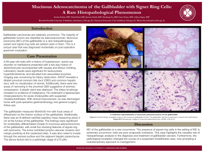
Figure: A collective representation of mucinous adenocarcinoma of the gallbladder.
Figure A: Gross findings of papillary mass on the mucosa of gallbladder fundus. The gallbladder wall has a mass that is mucoid with calcifications.
Figure B-D: Microscopic images of mucinous adenocarcinoma. x40 [B], x100 [C-D]
Figure A: Gross findings of papillary mass on the mucosa of gallbladder fundus. The gallbladder wall has a mass that is mucoid with calcifications.
Figure B-D: Microscopic images of mucinous adenocarcinoma. x40 [B], x100 [C-D]
Disclosures:
Swetha Paduri indicated no relevant financial relationships.
Nehal Patel indicated no relevant financial relationships.
Jessica Eisold indicated no relevant financial relationships.
Haoliang Xu indicated no relevant financial relationships.
Liam Nolan indicated no relevant financial relationships.
Aditya Gupta indicated no relevant financial relationships.
Swetha Paduri, MD1, Nehal Patel, MD1, Jessica Eisold, MD2, Haoliang Xu, MD3, Liam Nolan, MD3, Aditya Gupta, MD3. P0105 - Mucinous Adenocarcinoma of the Gallbladder with Signet Ring Cells: A Rare Histopathological Phenomenon, ACG 2023 Annual Scientific Meeting Abstracts. Vancouver, BC, Canada: American College of Gastroenterology.

