Sunday Poster Session
Category: Biliary/Pancreas
P0106 - Splenic Artery Aneurysm Disguised as a Pancreatic Mass
Sunday, October 22, 2023
3:30 PM - 7:00 PM PT
Location: Exhibit Hall

Has Audio

Jad Mitri, MD
St. Elizabeth's Medical Center
Boston, MA
Presenting Author(s)
Jad Mitri, MD1, Syed K. Mahmood, MD, MPH2
1St. Elizabeth's Medical Center, Boston, MA; 2St. Elizabeth's Medical Center, Brighton, MA
Introduction: Incidentally found pancreatic masses can often be challenging to define. Fine needle biopsy is indicated for masses showing high risk features on abdominal imaging. However, appropriate imaging protocols are necessary to avoid misdiagnosis.
Case Description/Methods: 54-year-old female had an incidentally found calcified mass in the tail of the pancreas concerning for malignancy on CT abdomen without contrast. Suspicion for malignancy was maintained on CT abdomen with contrast and MRI pancreas. On endoscopic ultrasound however, the mass turned out to be a splenic artery aneurysm. A fine needle biopsy of this mass could have resulted in life threatening bleeding complications.
Discussion: Careful evaluation of pancreatic masses is necessary prior to biopsy. CT contrast and MRI sensitivity for aneurysms may be insufficient, even when combined.

Disclosures:
Jad Mitri, MD1, Syed K. Mahmood, MD, MPH2. P0106 - Splenic Artery Aneurysm Disguised as a Pancreatic Mass, ACG 2023 Annual Scientific Meeting Abstracts. Vancouver, BC, Canada: American College of Gastroenterology.
1St. Elizabeth's Medical Center, Boston, MA; 2St. Elizabeth's Medical Center, Brighton, MA
Introduction: Incidentally found pancreatic masses can often be challenging to define. Fine needle biopsy is indicated for masses showing high risk features on abdominal imaging. However, appropriate imaging protocols are necessary to avoid misdiagnosis.
Case Description/Methods: 54-year-old female had an incidentally found calcified mass in the tail of the pancreas concerning for malignancy on CT abdomen without contrast. Suspicion for malignancy was maintained on CT abdomen with contrast and MRI pancreas. On endoscopic ultrasound however, the mass turned out to be a splenic artery aneurysm. A fine needle biopsy of this mass could have resulted in life threatening bleeding complications.
Discussion: Careful evaluation of pancreatic masses is necessary prior to biopsy. CT contrast and MRI sensitivity for aneurysms may be insufficient, even when combined.

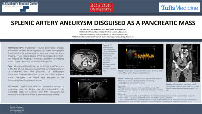
Figure: A: CT abdomen with contrast showing “a 2.0 cm intensely enhancing mass with rim-like calcifications in the tail of the pancreas. Adenocarcinoma cannot be excluded” (small red arrow).
B: Endoscopic ultrasound showing an anechoic lesion in the tail of pancreas with hyperechoic, shadowing calcifications in the wall of the lesion
C: Endoscopic ultrasound with Doppler showing brisk arterial flow within the pancreatic tail lesion
D: CT angiogram aorta with runoff coronal view confirming the mass as calcified splenic artery aneurysm measuring 2.6 x 2.7 cm (large thick arrow) with a clear connection to the splenic artery (small thick arrow)
Disclosures:
Jad Mitri indicated no relevant financial relationships.
Syed Mahmood indicated no relevant financial relationships.
Jad Mitri, MD1, Syed K. Mahmood, MD, MPH2. P0106 - Splenic Artery Aneurysm Disguised as a Pancreatic Mass, ACG 2023 Annual Scientific Meeting Abstracts. Vancouver, BC, Canada: American College of Gastroenterology.
