Sunday Poster Session
Category: Biliary/Pancreas
P0113 - A Rare Case of a Pancreaticopleural Fistula Presenting as Recurrent Bilateral Pleural Effusions
Sunday, October 22, 2023
3:30 PM - 7:00 PM PT
Location: Exhibit Hall

Has Audio
- AR
Abhinav Rao, MD
Trident Medical Center
Summerville, SC
Presenting Author(s)
Abhinav Rao, MD1, Fahim Syed, MD1, Angeli Patel, MD1, Brett VanLeer-Greenberg, MD2, James Ravenel, MD1, Julie Worthington, MD1
1Trident Medical Center, Charleston, SC; 2Trident Medical Center-HCA, Summerville, SC
Introduction: We present here a rare case of a young patient with recurrent bilateral pleural effusions found to have a pancreaticopleural fistula secondary to pseudocyst from chronic alcoholic pancreatitis.
Case Description/Methods: A 31-year-old male with a past medical history of chronic alcoholic pancreatitis, recent hospitalizations for alcoholic pancreatitis with pseudocyst formation and pleural effusions, and tobacco use presented to the emergency department for 1 day of worsening pleuritic chest pain.
His physical exam was remarkable for sinus tachycardia, bilaterally decreased breath sounds at lung bases, and left upper quadrant abdominal tenderness. Blood work was notable for hypokalemia of 3.2 mEq/L, leukocytosis of 11.8 k/mm3, hemoglobin 11.9 g/dL, thrombocytosis of 698 k/mm3, and a lipase of 159. CTAP revealed a minimally enlarging pancreatic pseudocyst within the proximal pancreatic body measuring 2.8 x 1.6 cm. CTA thorax showed increased and recurrent bilateral pleural effusions with a free-flowing large right-sided effusion, new large loculated effusion anteriorly, and moderate loculated left-sided effusion posteriorly.
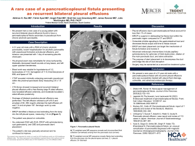
Drainage of bilateral exudative pleural effusions was performed. The left effusion showed a pH of 8.5 and amylase of 50,294 mcg/dL whereas the right effusion pH was 7.0 and amylase 187. Serological work up was unremarkable. Magnetic resonance cholangiopancreatography (MRCP) identified a fistulous tract extending from the body of the into the left pleural space, measuring 1.6 cm. (Figure 1A, 1B). The patient was initiated on octreotide therapy.
He underwent EGD with EUS, ERCP with sphincterotomy, cyst gastrostomy, and CBD and pancreatic stent placement. The patient’s diet was gradually advanced and he continued to improve.
Discussion: Pleural effusions due to pancreaticopleural fistula account for less than 1% of cases. MRCP is superior in delineating the fistula tract within the pancreatic region compared to CT and ERCP. Octreotide inhibits signaling to the gastrointestinal tract and significantly decreases fistula output and time to fistula closure. ERCP and stent placement can target the mechanism of fistula formation and reverse it; therapeutic interventions include papillary sphincterotomy for sphincter of Oddi dysfunction, dilatation of the main pancreatic duct, and stone extraction. The main purpose of the stent is to decompress the duct and bridge the site of duct disruption. Surgery may be warranted as a second line treatment option.

Disclosures:
Abhinav Rao, MD1, Fahim Syed, MD1, Angeli Patel, MD1, Brett VanLeer-Greenberg, MD2, James Ravenel, MD1, Julie Worthington, MD1. P0113 - A Rare Case of a Pancreaticopleural Fistula Presenting as Recurrent Bilateral Pleural Effusions, ACG 2023 Annual Scientific Meeting Abstracts. Vancouver, BC, Canada: American College of Gastroenterology.
1Trident Medical Center, Charleston, SC; 2Trident Medical Center-HCA, Summerville, SC
Introduction: We present here a rare case of a young patient with recurrent bilateral pleural effusions found to have a pancreaticopleural fistula secondary to pseudocyst from chronic alcoholic pancreatitis.
Case Description/Methods: A 31-year-old male with a past medical history of chronic alcoholic pancreatitis, recent hospitalizations for alcoholic pancreatitis with pseudocyst formation and pleural effusions, and tobacco use presented to the emergency department for 1 day of worsening pleuritic chest pain.
His physical exam was remarkable for sinus tachycardia, bilaterally decreased breath sounds at lung bases, and left upper quadrant abdominal tenderness. Blood work was notable for hypokalemia of 3.2 mEq/L, leukocytosis of 11.8 k/mm3, hemoglobin 11.9 g/dL, thrombocytosis of 698 k/mm3, and a lipase of 159. CTAP revealed a minimally enlarging pancreatic pseudocyst within the proximal pancreatic body measuring 2.8 x 1.6 cm. CTA thorax showed increased and recurrent bilateral pleural effusions with a free-flowing large right-sided effusion, new large loculated effusion anteriorly, and moderate loculated left-sided effusion posteriorly.
Drainage of bilateral exudative pleural effusions was performed. The left effusion showed a pH of 8.5 and amylase of 50,294 mcg/dL whereas the right effusion pH was 7.0 and amylase 187. Serological work up was unremarkable.
He underwent EGD with EUS, ERCP with sphincterotomy, cyst gastrostomy, and CBD and pancreatic stent placement. The patient’s diet was gradually advanced and he continued to improve.
Discussion: Pleural effusions due to pancreaticopleural fistula account for less than 1% of cases. MRCP is superior in delineating the fistula tract within the pancreatic region compared to CT and ERCP. Octreotide inhibits signaling to the gastrointestinal tract and significantly decreases fistula output and time to fistula closure. ERCP and stent placement can target the mechanism of fistula formation and reverse it; therapeutic interventions include papillary sphincterotomy for sphincter of Oddi dysfunction, dilatation of the main pancreatic duct, and stone extraction. The main purpose of the stent is to decompress the duct and bridge the site of duct disruption. Surgery may be warranted as a second line treatment option.

Figure: Figure 1. Pancreatico-pleural fistula.
A) T2 weighted axial MR sequence reveals well circumscribed fluid collection (arrowhead) arising from the pancreatic duct (arrows)
B) T2 weighted coronal MR sequence reveals fistula tract extending from the pancreas into the pleural space (arrows). E=pleural effusion. S= Stomach
A) T2 weighted axial MR sequence reveals well circumscribed fluid collection (arrowhead) arising from the pancreatic duct (arrows)
B) T2 weighted coronal MR sequence reveals fistula tract extending from the pancreas into the pleural space (arrows). E=pleural effusion. S= Stomach
Disclosures:
Abhinav Rao indicated no relevant financial relationships.
Fahim Syed indicated no relevant financial relationships.
Angeli Patel indicated no relevant financial relationships.
Brett VanLeer-Greenberg indicated no relevant financial relationships.
James Ravenel indicated no relevant financial relationships.
Julie Worthington indicated no relevant financial relationships.
Abhinav Rao, MD1, Fahim Syed, MD1, Angeli Patel, MD1, Brett VanLeer-Greenberg, MD2, James Ravenel, MD1, Julie Worthington, MD1. P0113 - A Rare Case of a Pancreaticopleural Fistula Presenting as Recurrent Bilateral Pleural Effusions, ACG 2023 Annual Scientific Meeting Abstracts. Vancouver, BC, Canada: American College of Gastroenterology.
