Sunday Poster Session
Category: Biliary/Pancreas
P0116 - Biliary Ductal Dilation Due to a Periampullary Diverticulum: It's Not Always a Stone
Sunday, October 22, 2023
3:30 PM - 7:00 PM PT
Location: Exhibit Hall

Has Audio

Samuel Purkey, DO
Allegheny Health Network
Pittsburgh, PA
Presenting Author(s)
Samuel Purkey, DO, Taimur S. Muzammil, MD, Rachel Toney, MD
Allegheny Health Network, Pittsburgh, PA
Introduction: Biliary ductal dilation is a common radiographic finding associated with right upper quadrant pain. Choledocholithiasis is a frequent cause of ductal dilation but what is the course of action when there is no radiographic stone present, but biliary obstruction is still suspected? We present a case of a periampullary diverticulum causing biliary ductal dilation associated with abdominal pain from chronic cholecystitis.
Case Description/Methods: We present a 31-year-old female with a 3 day history of right upper abdominal pain and nausea. Her liver function tests were normal but an abdominal ultrasound revealed gallbladder enlargement and wall thickening with common bile duct dilation to 9 mm. Contrasted CT of the abdomen also identified biliary ductal dilation to 7 mm with concern for a distal filling defect. ERCP was attempted but aborted due to difficulty cannulating the major papilla, as it was located within a duodenal diverticulum.
The patient was transferred to a high ERCP volume center. Given her normal bilirubin, an MRCP was pursued revealing common bile duct dilation to 7 mm and the presence of a periampullary diverticulum, which corresponded to the filling defect seen on prior CT imaging. There was no choledocholithiasis appreciated. Given the clinical presentation, it was felt that the patient's biliary ductal dilation was secondary to a periampullary diverticulum but not causing obstructive jaundice. Therefore, further ERCP attempts were discouraged. Surgery was consulted and she underwent a laparoscopic cholecystectomy with pathology consistent with chronic cholecystitis. She had resolution of her abdominal pain and was discharged with close follow-up.
Discussion: Biliary ductal dilation is a common radiographic finding associated with abdominal pain. When evaluating for choledocholithiasis, guidelines recommend classifying patients into high, intermediate, and low probability categories to aid clinical decision making. This patient was intermediate risk and MRCP imaging revealed the source of her biliary ductal dilation was a periampullary diverticulum. Therefore, repeat ERCP was not indicated. We present the abnormal case of a periampullary diverticulum presenting as incidental biliary ductal dilation in the setting of abdominal pain from chronic cholecystitis and not obstructive jaundice. This case demonstrates the importance of risk stratification of patients for whom there is suspicion for choledocholithiasis, as it is not always a stone and an ERCP may not be indicated.

Disclosures:
Samuel Purkey, DO, Taimur S. Muzammil, MD, Rachel Toney, MD. P0116 - Biliary Ductal Dilation Due to a Periampullary Diverticulum: It's Not Always a Stone, ACG 2023 Annual Scientific Meeting Abstracts. Vancouver, BC, Canada: American College of Gastroenterology.
Allegheny Health Network, Pittsburgh, PA
Introduction: Biliary ductal dilation is a common radiographic finding associated with right upper quadrant pain. Choledocholithiasis is a frequent cause of ductal dilation but what is the course of action when there is no radiographic stone present, but biliary obstruction is still suspected? We present a case of a periampullary diverticulum causing biliary ductal dilation associated with abdominal pain from chronic cholecystitis.
Case Description/Methods: We present a 31-year-old female with a 3 day history of right upper abdominal pain and nausea. Her liver function tests were normal but an abdominal ultrasound revealed gallbladder enlargement and wall thickening with common bile duct dilation to 9 mm. Contrasted CT of the abdomen also identified biliary ductal dilation to 7 mm with concern for a distal filling defect. ERCP was attempted but aborted due to difficulty cannulating the major papilla, as it was located within a duodenal diverticulum.
The patient was transferred to a high ERCP volume center. Given her normal bilirubin, an MRCP was pursued revealing common bile duct dilation to 7 mm and the presence of a periampullary diverticulum, which corresponded to the filling defect seen on prior CT imaging. There was no choledocholithiasis appreciated. Given the clinical presentation, it was felt that the patient's biliary ductal dilation was secondary to a periampullary diverticulum but not causing obstructive jaundice. Therefore, further ERCP attempts were discouraged. Surgery was consulted and she underwent a laparoscopic cholecystectomy with pathology consistent with chronic cholecystitis. She had resolution of her abdominal pain and was discharged with close follow-up.
Discussion: Biliary ductal dilation is a common radiographic finding associated with abdominal pain. When evaluating for choledocholithiasis, guidelines recommend classifying patients into high, intermediate, and low probability categories to aid clinical decision making. This patient was intermediate risk and MRCP imaging revealed the source of her biliary ductal dilation was a periampullary diverticulum. Therefore, repeat ERCP was not indicated. We present the abnormal case of a periampullary diverticulum presenting as incidental biliary ductal dilation in the setting of abdominal pain from chronic cholecystitis and not obstructive jaundice. This case demonstrates the importance of risk stratification of patients for whom there is suspicion for choledocholithiasis, as it is not always a stone and an ERCP may not be indicated.

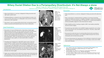
Figure: A: CT abdomen with contrast revealing biliary duct dilation and concern for a distal common bile duct filling defect (arrow).
B/C: MRCP imaging revealing biliary duct dilation to 7 mm and a 11 mm fluid filled duodenal diverticulum corresponding to the common bile duct filling defect seen on CT imaging.
D: MRCP imaging revealing biliary ductal dilation with no intraductal stone or filling defect present.
B/C: MRCP imaging revealing biliary duct dilation to 7 mm and a 11 mm fluid filled duodenal diverticulum corresponding to the common bile duct filling defect seen on CT imaging.
D: MRCP imaging revealing biliary ductal dilation with no intraductal stone or filling defect present.
Disclosures:
Samuel Purkey indicated no relevant financial relationships.
Taimur Muzammil indicated no relevant financial relationships.
Rachel Toney indicated no relevant financial relationships.
Samuel Purkey, DO, Taimur S. Muzammil, MD, Rachel Toney, MD. P0116 - Biliary Ductal Dilation Due to a Periampullary Diverticulum: It's Not Always a Stone, ACG 2023 Annual Scientific Meeting Abstracts. Vancouver, BC, Canada: American College of Gastroenterology.
