Sunday Poster Session
Category: Biliary/Pancreas
P0129 - Intra-Pancreatic Ectopic Adrenal Tissue Diagnosed on Endoscopic Ultrasound-Fine Needle Biopsy: First Report
Sunday, October 22, 2023
3:30 PM - 7:00 PM PT
Location: Exhibit Hall

Has Audio

Anusha Singhania, MBBS
Swedish Medical Center
Seattle, WA
Presenting Author(s)
Anusha Singhania, MBBS1, Tavishi Girotra, MBBS1, Shreya Chopra, MBBS1, Christopher J.. Carlson, MD1, Rajnish Mishra, MD1, Mohit Girotra, MD2
1Swedish Medical Center, Seattle, WA; 2Swedish Medical Center, Washington State University Elson S. Floyd School of Medicine, Seattle, WA
Introduction: Ectopic adrenal tissue is a benign condition, typically noted in organs along the migratory route of structures close to adrenal glands (gonads, kidneys), possibly due to abnormal cell adherence/migration during embryogenesis. When seen, it is usually in male children, on the right, usually asymptomatic and incidentally discovered during groin surgical exploration.
We describe a previously unreported entity of ectopic adrenal tissue in the tail of pancreas (TOP), in an elderly woman with no symptoms attributable to the abnormal mass.
Case Description/Methods: A 71-year old lady with HTN, hypothyroidism, old CVA, depression, renal cysts, chronic renal failure, alcohol-induced chronic pancreatitis (CP) underwent CT for surveillance of her left internal iliac artery aneurysm, which incidentally noted an 11x8 mm enhancing TOP lesion, with focal microcalcification.
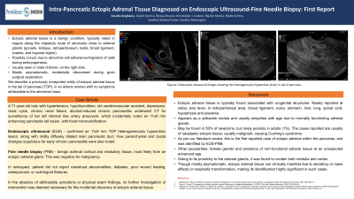
Patient was referred for endoscopic ultrasound (EUS), which confirmed an 11x9 mm TOP heterogeneously hypoechoic lesion, along with mildly diffusely dilated main pancreatic duct. Few parenchymal and ductal changes suspicious for early CP were also noted. Fine needle biopsy (FNB) revealed benign adrenal cortical and medullary tissue, most likely from an ectopic adrenal gland, negative for malignancy.
In retrospect, patient did not report menstrual abnormalities, diabetes, poor wound healing, osteoporosis, or cushingoid features. In the absence of attributable symptoms or physical exam findings, no further investigation or intervention was deemed necessary for this incidental discovery of ectopic adrenal tissue.
Discussion: Ectopic adrenal tissue is typically found associated with urogenital structures, and has rarely been reported at the celiac axis level, in retroperitoneal area, broad ligament, ovary, stomach, liver, lung, spinal cord, hypophysis and placenta. It appears as a yellowish nodule and usually atrophies with age due to normally functioning adrenal glands, very rarely persisting in adults, mostly in males.
Per our literature review, this is the first reported case of ectopic adrenal within the pancreas, identified by EUS-FNB. Other pecularities included female gender and presence of non-functional adrenal tissue at an unexpected advanced age. Owing to its proximity to the adrenal glands, it was found to contain both medulla and cortex. Though mostly asymptomatic, ectopic adrenal tissue can clinically manifest due to secretory or mass effects or neoplastic transformation, making its identification highly significant in such cases.

Disclosures:
Anusha Singhania, MBBS1, Tavishi Girotra, MBBS1, Shreya Chopra, MBBS1, Christopher J.. Carlson, MD1, Rajnish Mishra, MD1, Mohit Girotra, MD2. P0129 - Intra-Pancreatic Ectopic Adrenal Tissue Diagnosed on Endoscopic Ultrasound-Fine Needle Biopsy: First Report, ACG 2023 Annual Scientific Meeting Abstracts. Vancouver, BC, Canada: American College of Gastroenterology.
1Swedish Medical Center, Seattle, WA; 2Swedish Medical Center, Washington State University Elson S. Floyd School of Medicine, Seattle, WA
Introduction: Ectopic adrenal tissue is a benign condition, typically noted in organs along the migratory route of structures close to adrenal glands (gonads, kidneys), possibly due to abnormal cell adherence/migration during embryogenesis. When seen, it is usually in male children, on the right, usually asymptomatic and incidentally discovered during groin surgical exploration.
We describe a previously unreported entity of ectopic adrenal tissue in the tail of pancreas (TOP), in an elderly woman with no symptoms attributable to the abnormal mass.
Case Description/Methods: A 71-year old lady with HTN, hypothyroidism, old CVA, depression, renal cysts, chronic renal failure, alcohol-induced chronic pancreatitis (CP) underwent CT for surveillance of her left internal iliac artery aneurysm, which incidentally noted an 11x8 mm enhancing TOP lesion, with focal microcalcification.
Patient was referred for endoscopic ultrasound (EUS), which confirmed an 11x9 mm TOP heterogeneously hypoechoic lesion, along with mildly diffusely dilated main pancreatic duct. Few parenchymal and ductal changes suspicious for early CP were also noted. Fine needle biopsy (FNB) revealed benign adrenal cortical and medullary tissue, most likely from an ectopic adrenal gland, negative for malignancy.
In retrospect, patient did not report menstrual abnormalities, diabetes, poor wound healing, osteoporosis, or cushingoid features. In the absence of attributable symptoms or physical exam findings, no further investigation or intervention was deemed necessary for this incidental discovery of ectopic adrenal tissue.
Discussion: Ectopic adrenal tissue is typically found associated with urogenital structures, and has rarely been reported at the celiac axis level, in retroperitoneal area, broad ligament, ovary, stomach, liver, lung, spinal cord, hypophysis and placenta. It appears as a yellowish nodule and usually atrophies with age due to normally functioning adrenal glands, very rarely persisting in adults, mostly in males.
Per our literature review, this is the first reported case of ectopic adrenal within the pancreas, identified by EUS-FNB. Other pecularities included female gender and presence of non-functional adrenal tissue at an unexpected advanced age. Owing to its proximity to the adrenal glands, it was found to contain both medulla and cortex. Though mostly asymptomatic, ectopic adrenal tissue can clinically manifest due to secretory or mass effects or neoplastic transformation, making its identification highly significant in such cases.

Figure: EUS showing the heterogenously hypoechoic lesion in tail of pancreas
Disclosures:
Anusha Singhania indicated no relevant financial relationships.
Tavishi Girotra indicated no relevant financial relationships.
Shreya Chopra indicated no relevant financial relationships.
Christopher Carlson indicated no relevant financial relationships.
Rajnish Mishra indicated no relevant financial relationships.
Mohit Girotra indicated no relevant financial relationships.
Anusha Singhania, MBBS1, Tavishi Girotra, MBBS1, Shreya Chopra, MBBS1, Christopher J.. Carlson, MD1, Rajnish Mishra, MD1, Mohit Girotra, MD2. P0129 - Intra-Pancreatic Ectopic Adrenal Tissue Diagnosed on Endoscopic Ultrasound-Fine Needle Biopsy: First Report, ACG 2023 Annual Scientific Meeting Abstracts. Vancouver, BC, Canada: American College of Gastroenterology.
