Sunday Poster Session
Category: Biliary/Pancreas
P0133 - Klatskin Tumor Type III-B Presenting in an Elderly Patient with Concerning Abdominal Imaging
Sunday, October 22, 2023
3:30 PM - 7:00 PM PT
Location: Exhibit Hall

Has Audio

Alp S. Kahveci, MD
University of Iowa Hospital & Clinics
Iowa City, Iowa
Presenting Author(s)
Alp S. Kahveci, MD1, Jacob W. Cebulko, BS2, George Win, BS3, Syed Bilal Pasha, MD3, Faisal A.. Bukeirat, MD4
1University of Iowa Hospital & Clinics, Iowa City, IA; 2University of Missouri School of Medicine, Columbia, MO; 3University of Missouri-Columbia, Columbia, MO; 4University of Missouri at Columbia, Columbia, MO
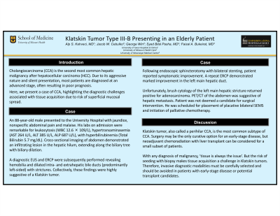
Introduction: Cholangiocarcinoma (CCA), the second most common hepatic malignancy after hepatocellular carcinoma (HCC), is a heterogenous tumor of the biliary system. Due to its aggressive nature and silent presentation, most patients are diagnosed at an advanced stage, often resulting in poor prognosis. Here, we present a case of CCA, highlighting the diagnostic challenges associated with tissue acquisition due to risk of superficial mucosal spread.
Case Description/Methods: An 88-year-old male presented to our institution with jaundice, nonspecific abdominal pain and malaise. His labs on admission were remarkable for leukocytosis (WBC 12.6 × 10⁹/L), elevated hepatic transaminases (AST 264 U/L, ALT 385 U/L, ALP 687 U/L) with hyperbilirubinemia (Total Bilirubin 5.7 mg/dL). Cross-sectional imaging of abdomen demonstrated an infiltrating lesion in the hepatic hilum, extending along the biliary tree with biliary dilation. Overall, his presentation was highly concerning for CCA.
A diagnostic endoscopic ultrasound (EUS) and endoscopic retrograde cholangiopancreatography (ERCP) were subsequently performed revealing hemobilia and dilated intra- and extrahepatic bile ducts (predominantly left-sided) with strictures (findings which were highly suggestive of Klatskin Tumor). Following endoscopic sphincterotomy with bilateral stenting, patient reported symptomatic improvement with repeat ERCP demonstrating marked improvement in the left main hepatic duct. Unfortunately, brush cytology of the left main hepatic stricture returned positive for adenocarcinoma. Meanwhile, PET/CT of the abdomen revealed concerning features for metastatic disease to the liver. Patient was not deemed a candidate for surgical intervention and was therefore scheduled for placement of placative bilateral SEMS and initiation of palliative chemotherapy.
Discussion: Anatomically, CCA is divided into three distinct subtypes, intrahepatic (iCCA), perihilar (pCCA; also called Klatskin tumor) and distal (dCCA). pCCA is the most common of these variants. Surgery may be the only curative option for an early-stage disease, but neoadjuvant chemoradiation with liver transplant can be considered for a small subset of patients with pCCA.
With any diagnosis of malignancy, ‘tissue is always the issue’. But the risk of seeding with biopsy makes tissue acquisition a challenge in Klatskin tumors. Therefore, invasive diagnostic modalities must be carefully selected and should be avoided in patients with early-stage disease or potential transplant candidates.
Disclosures:
Alp S. Kahveci, MD1, Jacob W. Cebulko, BS2, George Win, BS3, Syed Bilal Pasha, MD3, Faisal A.. Bukeirat, MD4. P0133 - Klatskin Tumor Type III-B Presenting in an Elderly Patient with Concerning Abdominal Imaging, ACG 2023 Annual Scientific Meeting Abstracts. Vancouver, BC, Canada: American College of Gastroenterology.
1University of Iowa Hospital & Clinics, Iowa City, IA; 2University of Missouri School of Medicine, Columbia, MO; 3University of Missouri-Columbia, Columbia, MO; 4University of Missouri at Columbia, Columbia, MO
Introduction: Cholangiocarcinoma (CCA), the second most common hepatic malignancy after hepatocellular carcinoma (HCC), is a heterogenous tumor of the biliary system. Due to its aggressive nature and silent presentation, most patients are diagnosed at an advanced stage, often resulting in poor prognosis. Here, we present a case of CCA, highlighting the diagnostic challenges associated with tissue acquisition due to risk of superficial mucosal spread.
Case Description/Methods: An 88-year-old male presented to our institution with jaundice, nonspecific abdominal pain and malaise. His labs on admission were remarkable for leukocytosis (WBC 12.6 × 10⁹/L), elevated hepatic transaminases (AST 264 U/L, ALT 385 U/L, ALP 687 U/L) with hyperbilirubinemia (Total Bilirubin 5.7 mg/dL). Cross-sectional imaging of abdomen demonstrated an infiltrating lesion in the hepatic hilum, extending along the biliary tree with biliary dilation. Overall, his presentation was highly concerning for CCA.
A diagnostic endoscopic ultrasound (EUS) and endoscopic retrograde cholangiopancreatography (ERCP) were subsequently performed revealing hemobilia and dilated intra- and extrahepatic bile ducts (predominantly left-sided) with strictures (findings which were highly suggestive of Klatskin Tumor). Following endoscopic sphincterotomy with bilateral stenting, patient reported symptomatic improvement with repeat ERCP demonstrating marked improvement in the left main hepatic duct. Unfortunately, brush cytology of the left main hepatic stricture returned positive for adenocarcinoma. Meanwhile, PET/CT of the abdomen revealed concerning features for metastatic disease to the liver. Patient was not deemed a candidate for surgical intervention and was therefore scheduled for placement of placative bilateral SEMS and initiation of palliative chemotherapy.
Discussion: Anatomically, CCA is divided into three distinct subtypes, intrahepatic (iCCA), perihilar (pCCA; also called Klatskin tumor) and distal (dCCA). pCCA is the most common of these variants. Surgery may be the only curative option for an early-stage disease, but neoadjuvant chemoradiation with liver transplant can be considered for a small subset of patients with pCCA.
With any diagnosis of malignancy, ‘tissue is always the issue’. But the risk of seeding with biopsy makes tissue acquisition a challenge in Klatskin tumors. Therefore, invasive diagnostic modalities must be carefully selected and should be avoided in patients with early-stage disease or potential transplant candidates.
Disclosures:
Alp Kahveci: Arbutus Biopharma Corp – Stock-publicly held company(excluding mutual/index funds). Bionano Genomics Inc – Stock-publicly held company(excluding mutual/index funds). Biontech SE – Stock-publicly held company(excluding mutual/index funds). Moderna Inc – Stock-publicly held company(excluding mutual/index funds). Pfizer Inc – Stock-publicly held company(excluding mutual/index funds). Senseonics Holdings Inc – Stock-publicly held company(excluding mutual/index funds).
Jacob Cebulko indicated no relevant financial relationships.
George Win indicated no relevant financial relationships.
Syed Bilal Pasha indicated no relevant financial relationships.
Faisal Bukeirat indicated no relevant financial relationships.
Alp S. Kahveci, MD1, Jacob W. Cebulko, BS2, George Win, BS3, Syed Bilal Pasha, MD3, Faisal A.. Bukeirat, MD4. P0133 - Klatskin Tumor Type III-B Presenting in an Elderly Patient with Concerning Abdominal Imaging, ACG 2023 Annual Scientific Meeting Abstracts. Vancouver, BC, Canada: American College of Gastroenterology.

