Sunday Poster Session
Category: Colon
P0234 - Incidental Finding of an Appendiceal Abscess on Screening Colonoscopy
Sunday, October 22, 2023
3:30 PM - 7:00 PM PT
Location: Exhibit Hall

Has Audio
.jpg)
Joseph David, MD
University of Arizona College of Medicine
Phoenix, AZ
Presenting Author(s)
Brian M.. Fung, MD, Joseph David, MD
University of Arizona College of Medicine, Phoenix, AZ
Introduction: Acute appendicitis, typically presenting with right lower-quadrant abdominal pain, nausea, and vomiting, is a relatively common infection, with an overall estimated lifetime risk of approximately 7-8%. Occult appendicitis (appendicitis without symptoms), on the other hand, is extremely rare. In the following case, we describe the incidental diagnosis and management of an appendiceal abscess found on screening colonoscopy.
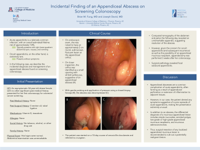
Case Description/Methods: An asymptomatic 54-year-old obese female with no other significant past medical history presented for her first colonoscopy for colorectal cancer screening. She denied having abdominal pain, rectal bleeding, or changes in bowel habits. Her past surgical history was significant for three cesarean sections and tubal ligation. She denied tobacco or alcohol use, and her only medications were vitamin D and trazodone for sleep. She denied a family history of gastrointestinal issues. On endoscopic exam, she was noted to have an approximately 2 cm round, submucosal, fluctuant lesion at the appendiceal orifice. On closer inspection, the orifice was identified as a small opening with whitish yellow pus, suggestive of an appendiceal abscess (Figure 1). With gentle probing and application of pressure using a closed biopsy forceps, the abscess was decompressed. The patient was started on a 10-day course of amoxicillin-clavulanate and referred to a surgeon. Computed tomography of the abdomen and pelvis the following day revealed an unremarkable appendix, suggesting resolution of the abscess. However, given the concern for occult appendicitis (and subsequent recurrence) as well as the possibility of an appendiceal mucinous neoplasm, appendectomy was performed 3 weeks after her colonoscopy. Surgical pathology revealed focal subacute appendicitis.
Discussion: Appendiceal abscesses are a common complication of acute appendicitis, often forming as a result of appendiceal perforation or extension of inflammation to adjacent tissues. However, in our case, the patient denied any symptoms suggestive of a prior episode of acute appendicitis, making the presentation extremely unusual. In addition to an abscess, the differential diagnosis of a mucinous appendiceal lesion includes simple mucoceles, serrated polyps, low-grade or high-grade appendiceal mucinous neoplasms, and mucinous adenocarcinomas; thus, surgical resection of any localized appendiceal mucinous lesion is recommended to rule out a potentially malignant lesion.

Disclosures:
Brian M.. Fung, MD, Joseph David, MD. P0234 - Incidental Finding of an Appendiceal Abscess on Screening Colonoscopy, ACG 2023 Annual Scientific Meeting Abstracts. Vancouver, BC, Canada: American College of Gastroenterology.
University of Arizona College of Medicine, Phoenix, AZ
Introduction: Acute appendicitis, typically presenting with right lower-quadrant abdominal pain, nausea, and vomiting, is a relatively common infection, with an overall estimated lifetime risk of approximately 7-8%. Occult appendicitis (appendicitis without symptoms), on the other hand, is extremely rare. In the following case, we describe the incidental diagnosis and management of an appendiceal abscess found on screening colonoscopy.
Case Description/Methods: An asymptomatic 54-year-old obese female with no other significant past medical history presented for her first colonoscopy for colorectal cancer screening. She denied having abdominal pain, rectal bleeding, or changes in bowel habits. Her past surgical history was significant for three cesarean sections and tubal ligation. She denied tobacco or alcohol use, and her only medications were vitamin D and trazodone for sleep. She denied a family history of gastrointestinal issues. On endoscopic exam, she was noted to have an approximately 2 cm round, submucosal, fluctuant lesion at the appendiceal orifice. On closer inspection, the orifice was identified as a small opening with whitish yellow pus, suggestive of an appendiceal abscess (Figure 1). With gentle probing and application of pressure using a closed biopsy forceps, the abscess was decompressed. The patient was started on a 10-day course of amoxicillin-clavulanate and referred to a surgeon. Computed tomography of the abdomen and pelvis the following day revealed an unremarkable appendix, suggesting resolution of the abscess. However, given the concern for occult appendicitis (and subsequent recurrence) as well as the possibility of an appendiceal mucinous neoplasm, appendectomy was performed 3 weeks after her colonoscopy. Surgical pathology revealed focal subacute appendicitis.
Discussion: Appendiceal abscesses are a common complication of acute appendicitis, often forming as a result of appendiceal perforation or extension of inflammation to adjacent tissues. However, in our case, the patient denied any symptoms suggestive of a prior episode of acute appendicitis, making the presentation extremely unusual. In addition to an abscess, the differential diagnosis of a mucinous appendiceal lesion includes simple mucoceles, serrated polyps, low-grade or high-grade appendiceal mucinous neoplasms, and mucinous adenocarcinomas; thus, surgical resection of any localized appendiceal mucinous lesion is recommended to rule out a potentially malignant lesion.

Figure: A protruding appendiceal lesion with white pus noted on colonoscopy exam.
Disclosures:
Brian Fung indicated no relevant financial relationships.
Joseph David indicated no relevant financial relationships.
Brian M.. Fung, MD, Joseph David, MD. P0234 - Incidental Finding of an Appendiceal Abscess on Screening Colonoscopy, ACG 2023 Annual Scientific Meeting Abstracts. Vancouver, BC, Canada: American College of Gastroenterology.
