Sunday Poster Session
Category: Colon
P0253 - A Rare Case of Rectal Mucinous Adenocarcinoma in Quiescent Crohn's Disease
Sunday, October 22, 2023
3:30 PM - 7:00 PM PT
Location: Exhibit Hall

Has Audio

Christine Garabetian, MD
West Anaheim Medical Center
Anaheim, CA
Presenting Author(s)
Christine Garabetian, MD, Amit Bhanvadia, MD
West Anaheim Medical Center, Anaheim, CA
Introduction: More uncommon than mucinous colorectal cancer (CRC) is localized rectal mucinous adenocarcinoma, a distinct histopathological subtype of rectal cancer. Key features of mucinous adenocarcinomas (MACs) are aberrant and abundant mucin expression and composition, which can be seen on MRI. Rectal MAC, found in less than 20% of CRC patients, is associated with genetic aberrations resulting in an aggressive and therapy-resistant cancer. Those with inflammatory bowel disease (IBD) such as Crohn’s disease (CD) have higher prevalence of CRC, and colorectal MAC, than the general population due to chronic inflammation causing continuous cell turnover within the intestinal tract leading to increased risk of mutations.
Case Description/Methods: A 57-year-old male with a 25-year-history of well-managed CD and without current active disease presented for routine yearly colonoscopy. Colonoscopy done a year prior was only remarkable for internal hemorrhoids. Colonic and rectal biopsies at the time were consistent with quiescent disease without evidence of dysplasia or neoplasia.
Colonoscopy during this admission showed no endoscopic or histopathological evidence of CD in the colon or rectum. Internal hemorrhoids were noted as well as a circumferential, ulcerated, hard and nodular lesion proximal to the dentate line in the rectum (Figure A). Rectal biopsy histopathology revealed invasive adenocarcinoma, with the tumor showing infiltrating clusters of tumor cells with abundant mucus production.
Pelvic MRI demonstrated a 3.2 x 2.6 x 1.6 cm mucinous mass (Figure B-D) involving the right rectum, and appearing to be contained within the rectum superior to the anus. Staging CT demonstrated moderate-sized rectal mass consistent with neoplasm but negative for metastatic disease. Diagnosis was stage IIA (cT3, cN0, cM0) rectal MAC. Patient has started neoadjuvant chemoradiation therapy with plans for surgical resection.
Discussion: Colorectal MAC occurs most commonly in younger females and is predominantly located in the proximal colon; localized rectal tumor is very rare. Compared to its non-mucinous counterpart, rectal MACs are diagnosed at a higher T category, have worse prognosis, and greater risk of metastases. Factors involved in MAC development are unclear, but cases and identifications of conditions associated with MAC, specifically rectal MAC, can pave the way for multidisciplinary patient care and treatment modalities while providing insight to the etiological pathways which lead to this rare cancer subtype.

Disclosures:
Christine Garabetian, MD, Amit Bhanvadia, MD. P0253 - A Rare Case of Rectal Mucinous Adenocarcinoma in Quiescent Crohn's Disease, ACG 2023 Annual Scientific Meeting Abstracts. Vancouver, BC, Canada: American College of Gastroenterology.
West Anaheim Medical Center, Anaheim, CA
Introduction: More uncommon than mucinous colorectal cancer (CRC) is localized rectal mucinous adenocarcinoma, a distinct histopathological subtype of rectal cancer. Key features of mucinous adenocarcinomas (MACs) are aberrant and abundant mucin expression and composition, which can be seen on MRI. Rectal MAC, found in less than 20% of CRC patients, is associated with genetic aberrations resulting in an aggressive and therapy-resistant cancer. Those with inflammatory bowel disease (IBD) such as Crohn’s disease (CD) have higher prevalence of CRC, and colorectal MAC, than the general population due to chronic inflammation causing continuous cell turnover within the intestinal tract leading to increased risk of mutations.
Case Description/Methods: A 57-year-old male with a 25-year-history of well-managed CD and without current active disease presented for routine yearly colonoscopy. Colonoscopy done a year prior was only remarkable for internal hemorrhoids. Colonic and rectal biopsies at the time were consistent with quiescent disease without evidence of dysplasia or neoplasia.
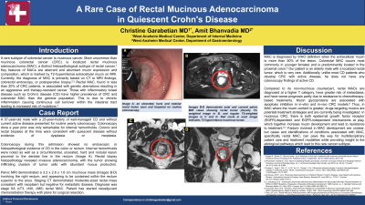
Colonoscopy during this admission showed no endoscopic or histopathological evidence of CD in the colon or rectum. Internal hemorrhoids were noted as well as a circumferential, ulcerated, hard and nodular lesion proximal to the dentate line in the rectum (Figure A). Rectal biopsy histopathology revealed invasive adenocarcinoma, with the tumor showing infiltrating clusters of tumor cells with abundant mucus production.
Pelvic MRI demonstrated a 3.2 x 2.6 x 1.6 cm mucinous mass (Figure B-D) involving the right rectum, and appearing to be contained within the rectum superior to the anus. Staging CT demonstrated moderate-sized rectal mass consistent with neoplasm but negative for metastatic disease. Diagnosis was stage IIA (cT3, cN0, cM0) rectal MAC. Patient has started neoadjuvant chemoradiation therapy with plans for surgical resection.
Discussion: Colorectal MAC occurs most commonly in younger females and is predominantly located in the proximal colon; localized rectal tumor is very rare. Compared to its non-mucinous counterpart, rectal MACs are diagnosed at a higher T category, have worse prognosis, and greater risk of metastases. Factors involved in MAC development are unclear, but cases and identifications of conditions associated with MAC, specifically rectal MAC, can pave the way for multidisciplinary patient care and treatment modalities while providing insight to the etiological pathways which lead to this rare cancer subtype.

Figure: Image A shows an ulcerated, hard, and nodular rectal lesion seen and biopsied on routine colonoscopy. Images B-D demonstrate axial and coronal pelvic MRI views showing rectal tumor (heavily T2-weighted image in B, and regular T2-weighted images in C and D. Red circle in each image indicates T2-hyperintense mucinous tumor.
Disclosures:
Christine Garabetian indicated no relevant financial relationships.
Amit Bhanvadia indicated no relevant financial relationships.
Christine Garabetian, MD, Amit Bhanvadia, MD. P0253 - A Rare Case of Rectal Mucinous Adenocarcinoma in Quiescent Crohn's Disease, ACG 2023 Annual Scientific Meeting Abstracts. Vancouver, BC, Canada: American College of Gastroenterology.
