Sunday Poster Session
Category: Colon
P0290 - Adenocarcinoma of Colon Presenting as a Mixed Cystic-Solid Adnexal Mass Causing Sigmoid Colon Stricture
Sunday, October 22, 2023
3:30 PM - 7:00 PM PT
Location: Exhibit Hall

Has Audio

Jerapas Thongpiya, MD
Texas Tech University Health Sciences Center
Lubbock, TX
Presenting Author(s)
Jerapas Thongpiya, MD1, Sakditad Saowapa, MD1, Pitchaporn Yingchoncharoen, MD1, Arif Daoud, MD1, Boyd Fenton, MD2, Sameer Islam, MD, MBA1
1Texas Tech University Health Sciences Center, Lubbock, TX; 2University Medical Center, Lubbock, TX
Introduction: Adnexal masses represent a spectrum of conditions from a gynecologic or non-gynecologic source and benign or malignant. The most common symptoms are pelvic or abdominal pain, distention, bloating, intestinal obstruction, or lower urinary tract symptoms. Here, we present an unusual presentation of adenocarcinoma of colon cancer presenting with solid/cystic adnexal mass causing extrinsic luminal compression of the sigmoid colon.
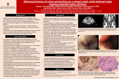
Case Description/Methods: A 61-year-old woman with a past medical history of COPD, hypertension, hypothyroidism, CAD s/p stents placement, heavy smoking and drinking presented with progressive LLQ abdominal pain for 3 months. Physical examination showed LLQ tenderness. Labs showed elevated CEA 29.6ng/mL (normal < =3.8) otherwise negative other tumor markers. Her family history was significant for colon cancer in her brother. CT abdomen showed a complex solid/cystic mass in the left adnexal area (10cm of solid and 6cm of cystic components) with abutment and possible invasion to sigmoid colon. Colonoscopy was performed and revealed stricture without typical appearance of colon cancer in the sigmoid colon at 50cm from anus, which caused near complete obstruction that scope couldn’t traverse. Her pathology from stricture biopsy showed invasive, moderately differentiated adenocarcinoma with colorectal origin from positive SATB-2 and negative PAX-8, which was consistent with her elevated CEA. She underwent exploratory laparotomy with Hartman's procedure, small bowel resection anastomosis, left SO, and ureteral stent, which revealed pT4bN0M0 stage from final pathology result. Her hospital course was complicated by secondary peritonitis but improved with antibiotics.
Discussion: Colorectal cancer is common and one of the three leading causes of death in women. Cancers of the left and sigmoid colon are often deeply invasive and annular, patients can be asymptomatic or present with abdominal pain, bowel habit change or obstructive symptoms. Adnexal masses especially in postmenopausal women raise concerns of malignancy which necessitates thorough evaluation. Tumor markers may aid in identifying carcinoma origin but are not sensitive. In this case, the patient had elevated CEA with a mixed solid cystic adnexal mass and was proven to have adenocarcinoma of the sigmoid colon causing intraluminal compression as opposed to gynecologic malignancy. Patients with pelvic masses should receive careful evaluation including applicable endoscopic studies and to minimize the risks of misdiagnosis.

Disclosures:
Jerapas Thongpiya, MD1, Sakditad Saowapa, MD1, Pitchaporn Yingchoncharoen, MD1, Arif Daoud, MD1, Boyd Fenton, MD2, Sameer Islam, MD, MBA1. P0290 - Adenocarcinoma of Colon Presenting as a Mixed Cystic-Solid Adnexal Mass Causing Sigmoid Colon Stricture, ACG 2023 Annual Scientific Meeting Abstracts. Vancouver, BC, Canada: American College of Gastroenterology.
1Texas Tech University Health Sciences Center, Lubbock, TX; 2University Medical Center, Lubbock, TX
Introduction: Adnexal masses represent a spectrum of conditions from a gynecologic or non-gynecologic source and benign or malignant. The most common symptoms are pelvic or abdominal pain, distention, bloating, intestinal obstruction, or lower urinary tract symptoms. Here, we present an unusual presentation of adenocarcinoma of colon cancer presenting with solid/cystic adnexal mass causing extrinsic luminal compression of the sigmoid colon.
Case Description/Methods: A 61-year-old woman with a past medical history of COPD, hypertension, hypothyroidism, CAD s/p stents placement, heavy smoking and drinking presented with progressive LLQ abdominal pain for 3 months. Physical examination showed LLQ tenderness. Labs showed elevated CEA 29.6ng/mL (normal < =3.8) otherwise negative other tumor markers. Her family history was significant for colon cancer in her brother. CT abdomen showed a complex solid/cystic mass in the left adnexal area (10cm of solid and 6cm of cystic components) with abutment and possible invasion to sigmoid colon. Colonoscopy was performed and revealed stricture without typical appearance of colon cancer in the sigmoid colon at 50cm from anus, which caused near complete obstruction that scope couldn’t traverse. Her pathology from stricture biopsy showed invasive, moderately differentiated adenocarcinoma with colorectal origin from positive SATB-2 and negative PAX-8, which was consistent with her elevated CEA. She underwent exploratory laparotomy with Hartman's procedure, small bowel resection anastomosis, left SO, and ureteral stent, which revealed pT4bN0M0 stage from final pathology result. Her hospital course was complicated by secondary peritonitis but improved with antibiotics.
Discussion: Colorectal cancer is common and one of the three leading causes of death in women. Cancers of the left and sigmoid colon are often deeply invasive and annular, patients can be asymptomatic or present with abdominal pain, bowel habit change or obstructive symptoms. Adnexal masses especially in postmenopausal women raise concerns of malignancy which necessitates thorough evaluation. Tumor markers may aid in identifying carcinoma origin but are not sensitive. In this case, the patient had elevated CEA with a mixed solid cystic adnexal mass and was proven to have adenocarcinoma of the sigmoid colon causing intraluminal compression as opposed to gynecologic malignancy. Patients with pelvic masses should receive careful evaluation including applicable endoscopic studies and to minimize the risks of misdiagnosis.

Figure: CT abdomen showed a complex solid/cystic mass in the left adnexal area (10cm of solid and 6cm of cystic components) with abutment and possible invasion to sigmoid colon.
Colonoscopy showed stricture without typical appearance of colon cancer in the sigmoid colon at 50cm from anus causing near complete obstruction that scope couldn't traverse.
SATB-2 image showed positive staining in the glands, supporting colorectal primary
H&E 10x image showed infiltrating malignant glands with associated desmoplastic reaction (invasive colon adenocarcinoma)
Disclosures:
Jerapas Thongpiya indicated no relevant financial relationships.
Sakditad Saowapa indicated no relevant financial relationships.
Pitchaporn Yingchoncharoen indicated no relevant financial relationships.
Arif Daoud indicated no relevant financial relationships.
Boyd Fenton indicated no relevant financial relationships.
Sameer Islam indicated no relevant financial relationships.
Jerapas Thongpiya, MD1, Sakditad Saowapa, MD1, Pitchaporn Yingchoncharoen, MD1, Arif Daoud, MD1, Boyd Fenton, MD2, Sameer Islam, MD, MBA1. P0290 - Adenocarcinoma of Colon Presenting as a Mixed Cystic-Solid Adnexal Mass Causing Sigmoid Colon Stricture, ACG 2023 Annual Scientific Meeting Abstracts. Vancouver, BC, Canada: American College of Gastroenterology.
