Sunday Poster Session
Category: Esophagus
P0441 - Dysphagia From Lymphocytic Esophagitis Successfully Treated With Proton Pump Inhibitor
Sunday, October 22, 2023
3:30 PM - 7:00 PM PT
Location: Exhibit Hall

Has Audio

Aidan J. David
Case Western Reserve University
Cleveland, OH
Presenting Author(s)
Aidan J. David, 1, Brian M.. Fung, MD2, Joseph David, MD2
1Case Western Reserve University, Cleveland, OH; 2University of Arizona College of Medicine, Phoenix, AZ
Introduction: Lymphocytic esophagitis (LE) is a rare condition seen in 0.1% of all esophageal biopsies in the United States. It is characterized by a large number of intracellular lymphocytes in the esophageal mucosa with relative paucity of other inflammatory cells. The most common symptom associated with LE is dysphagia, and endoscopic findings can vary from normal-appearing mucosa to features such as furrows, rings, and stenosis, all of which are findings observed in the more commonly diagnosed condition, eosinophilic esophagitis (EoE). Here, we present a case of dysphagia secondary to LE in which a proton pump inhibitor (PPI) was used to alleviate symptoms.
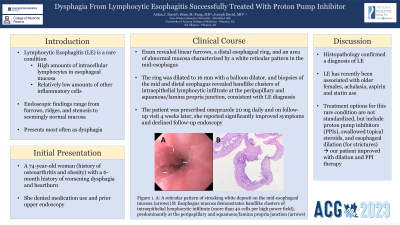
Case Description/Methods: A 74-year-old woman with a history of osteoarthritis and obesity presented to the clinic with a 6 month history of worsening dysphagia to meat and bread. She also reported worsening heartburn in her lower sternum after drinking carbonated beverages and white wine. She denied taking any medications and never had a prior upper endoscopy. On endoscopic examination, she was noted to have linear furrows, a distal esophageal ring, and an area of abnormal mucosa characterized by a white reticular pattern in the mid-esophagus (Figure 1 Panel A). The ring was dilated with a 16 mm balloon, and biopsies of the mid- and distal esophagus revealed bandlike clusters of intraepithelial lymphocytic infiltrate at a density of more than 40 cells per high power field, predominantly at the peripapillary and squamous/lamina propria junction, consistent with a diagnosis of lymphocytic esophagitis (Figure 1 Panel B). The patient was prescribed omeprazole 20 mg daily. On follow-up visit 4 weeks later, she reported significantly improved symptoms and declined follow-up endoscopy.
Discussion: Dysphagia is a common complaint seen in the gastroenterology clinic. Anywhere between 12-22% of patients with dysphagia are diagnosed with EoE. In our patient, symptoms and endoscopic findings were mostly suggestive of EoE (linear furrows and an esophageal ring); however, a reticular mucosal pattern was also observed, which is uncharacteristic of EoE. Histopathology ultimately confirmed a diagnosis of LE, a distinct entity that is significantly rarer. LE has recently been associated with older females, achalasia, aspirin and statin use. Treatment options for this condition are not standardized, but include PPIs, swallowed topical steroids, and esophageal dilation (for strictures); our patient improved with dilation and PPI therapy.

Disclosures:
Aidan J. David, 1, Brian M.. Fung, MD2, Joseph David, MD2. P0441 - Dysphagia From Lymphocytic Esophagitis Successfully Treated With Proton Pump Inhibitor, ACG 2023 Annual Scientific Meeting Abstracts. Vancouver, BC, Canada: American College of Gastroenterology.
1Case Western Reserve University, Cleveland, OH; 2University of Arizona College of Medicine, Phoenix, AZ
Introduction: Lymphocytic esophagitis (LE) is a rare condition seen in 0.1% of all esophageal biopsies in the United States. It is characterized by a large number of intracellular lymphocytes in the esophageal mucosa with relative paucity of other inflammatory cells. The most common symptom associated with LE is dysphagia, and endoscopic findings can vary from normal-appearing mucosa to features such as furrows, rings, and stenosis, all of which are findings observed in the more commonly diagnosed condition, eosinophilic esophagitis (EoE). Here, we present a case of dysphagia secondary to LE in which a proton pump inhibitor (PPI) was used to alleviate symptoms.
Case Description/Methods: A 74-year-old woman with a history of osteoarthritis and obesity presented to the clinic with a 6 month history of worsening dysphagia to meat and bread. She also reported worsening heartburn in her lower sternum after drinking carbonated beverages and white wine. She denied taking any medications and never had a prior upper endoscopy. On endoscopic examination, she was noted to have linear furrows, a distal esophageal ring, and an area of abnormal mucosa characterized by a white reticular pattern in the mid-esophagus (Figure 1 Panel A). The ring was dilated with a 16 mm balloon, and biopsies of the mid- and distal esophagus revealed bandlike clusters of intraepithelial lymphocytic infiltrate at a density of more than 40 cells per high power field, predominantly at the peripapillary and squamous/lamina propria junction, consistent with a diagnosis of lymphocytic esophagitis (Figure 1 Panel B). The patient was prescribed omeprazole 20 mg daily. On follow-up visit 4 weeks later, she reported significantly improved symptoms and declined follow-up endoscopy.
Discussion: Dysphagia is a common complaint seen in the gastroenterology clinic. Anywhere between 12-22% of patients with dysphagia are diagnosed with EoE. In our patient, symptoms and endoscopic findings were mostly suggestive of EoE (linear furrows and an esophageal ring); however, a reticular mucosal pattern was also observed, which is uncharacteristic of EoE. Histopathology ultimately confirmed a diagnosis of LE, a distinct entity that is significantly rarer. LE has recently been associated with older females, achalasia, aspirin and statin use. Treatment options for this condition are not standardized, but include PPIs, swallowed topical steroids, and esophageal dilation (for strictures); our patient improved with dilation and PPI therapy.

Figure: Figure 1. A: A reticular pattern of streaking white deposit on the mid-esophageal mucosa. B: Esophagus mucosa demonstrates bandlike clusters of intraepithelial lymphocytic infiltrate (more than 40 cells per high power field), predominantly at the peripapillary and squamous/lamina propria junction.
Disclosures:
Aidan David indicated no relevant financial relationships.
Brian Fung indicated no relevant financial relationships.
Joseph David indicated no relevant financial relationships.
Aidan J. David, 1, Brian M.. Fung, MD2, Joseph David, MD2. P0441 - Dysphagia From Lymphocytic Esophagitis Successfully Treated With Proton Pump Inhibitor, ACG 2023 Annual Scientific Meeting Abstracts. Vancouver, BC, Canada: American College of Gastroenterology.
