Sunday Poster Session
Category: Esophagus
P0463 - An Atypical Case of Esophageal Abrikossoff's Tumor in a Background of Barrett’s Esophagus: A Coincidence?
Sunday, October 22, 2023
3:30 PM - 7:00 PM PT
Location: Exhibit Hall

Has Audio
.jpg)
Jiteshwar S. Pannu, MD
University of Oklahoma Health Sciences Center
Oklahoma City, OK
Presenting Author(s)
Jiteshwar S. Pannu, MD, Nancy T Nguyen, , Shruti Mony, MD, Niveditha Popuri, MD
University of Oklahoma Health Sciences Center, Oklahoma City, OK
Introduction: Esophageal granular cell tumors (GCTs) aka Abrikossoff's tumors are rare neuroectodermal neoplasms, incidentally discovered during esophagogastroduodenoscopy (EGD). While majority of them are benign, 1-2% of GCTs are malignant. There is no known association of GCTs with other esophageal disorders. We present a unique case of esophageal GCT, in the setting of long segment Barrett’s esophagus (BE).
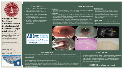
Case Description/Methods: A 62-year-old male with a history of chronic GERD, long segment BE with low grade dysplasia, underwent an outpatient surveillance EGD. In addition to long segment BE, classified as C5-M6 per Prague criteria, a 1 cm sub-epithelial nodule was seen 40 cm from the incisors in the distal esophagus, within the Barrett’s segment (Figure A/B).
An EUS was performed, which confirmed the round, homogeneous nodule arising from the muscularis mucosal layer, measuring 1 cm in size (Figure C). Given the unclear etiology of the lesion in a background of dysplasia, decision was made for endoscopic resection. The nodule was successfully resected via en bloc endoscopic mucosal resection (EMR) (Figure D). Histopathological analysis was consistent with GCT and intestinal/goblet cell metaplasia indicative of BE, and no evidence of dysplasia (Figure E). Immunohistochemistry (IHC) revealed diffuse S100 immunoreactivity, hence confirming the diagnosis (Figure F). There were no post procedural complications. Patient is scheduled for endoscopic surveillance in 6 months.
Discussion: Gastrointestinal GCTs account for 8% of all GCTs, of which a third are found incidentally in the esophagus. During EGD, they characteristically appear as round, well-defined, yellow-white nodules with a firm-hard consistency and normal overlying mucosa. EUS aids in differentiating GCTs from other sub-epithelial lesions. Classic histopathology and IHC are indispensable in confirming the diagnosis, with positive S100 being a reliable marker of Schwann cell origin. The optimal management of GCTs is still controversial, but endoscopic resection is recommended for symptomatic tumors greater than 1 cm and GCTs with atypical ultrasonographic or pathologic features, with surgery being reserved for larger tumors. There is no known association between BE and GCTs, and a causal relationship has not been hypothesized in the literature before. In the setting of BE, GCTs can present with other neoplasms such as leiomyomas and intra-mucosal adenocarcinomas, hence surveillance and timely resection should be practiced.

Disclosures:
Jiteshwar S. Pannu, MD, Nancy T Nguyen, , Shruti Mony, MD, Niveditha Popuri, MD. P0463 - An Atypical Case of Esophageal Abrikossoff's Tumor in a Background of Barrett’s Esophagus: A Coincidence?, ACG 2023 Annual Scientific Meeting Abstracts. Vancouver, BC, Canada: American College of Gastroenterology.
University of Oklahoma Health Sciences Center, Oklahoma City, OK
Introduction: Esophageal granular cell tumors (GCTs) aka Abrikossoff's tumors are rare neuroectodermal neoplasms, incidentally discovered during esophagogastroduodenoscopy (EGD). While majority of them are benign, 1-2% of GCTs are malignant. There is no known association of GCTs with other esophageal disorders. We present a unique case of esophageal GCT, in the setting of long segment Barrett’s esophagus (BE).
Case Description/Methods: A 62-year-old male with a history of chronic GERD, long segment BE with low grade dysplasia, underwent an outpatient surveillance EGD. In addition to long segment BE, classified as C5-M6 per Prague criteria, a 1 cm sub-epithelial nodule was seen 40 cm from the incisors in the distal esophagus, within the Barrett’s segment (Figure A/B).
An EUS was performed, which confirmed the round, homogeneous nodule arising from the muscularis mucosal layer, measuring 1 cm in size (Figure C). Given the unclear etiology of the lesion in a background of dysplasia, decision was made for endoscopic resection. The nodule was successfully resected via en bloc endoscopic mucosal resection (EMR) (Figure D). Histopathological analysis was consistent with GCT and intestinal/goblet cell metaplasia indicative of BE, and no evidence of dysplasia (Figure E). Immunohistochemistry (IHC) revealed diffuse S100 immunoreactivity, hence confirming the diagnosis (Figure F). There were no post procedural complications. Patient is scheduled for endoscopic surveillance in 6 months.
Discussion: Gastrointestinal GCTs account for 8% of all GCTs, of which a third are found incidentally in the esophagus. During EGD, they characteristically appear as round, well-defined, yellow-white nodules with a firm-hard consistency and normal overlying mucosa. EUS aids in differentiating GCTs from other sub-epithelial lesions. Classic histopathology and IHC are indispensable in confirming the diagnosis, with positive S100 being a reliable marker of Schwann cell origin. The optimal management of GCTs is still controversial, but endoscopic resection is recommended for symptomatic tumors greater than 1 cm and GCTs with atypical ultrasonographic or pathologic features, with surgery being reserved for larger tumors. There is no known association between BE and GCTs, and a causal relationship has not been hypothesized in the literature before. In the setting of BE, GCTs can present with other neoplasms such as leiomyomas and intra-mucosal adenocarcinomas, hence surveillance and timely resection should be practiced.

Figure: Figure A : demonstrates the 1 cm sub-epithelial nodule (yellow arrow) seen in the distal esophagus, within the Barrett's segment;
Figure B : shows the 'tongue-like' extensions above the gastroesophageal junction with an irregular Z line (red arrowheads) indicative of Barrett's esophagus with distally visible sub-epithelial nodule (yellow arrow);
Figure C : EUS showing the 1 cm round, homogenous, sub-epithelial nodule (yellow arrow) arising from the muscularis mucosal layer;
Figure D : En bloc endoscopic mucosal resection of the sub-epithelial nodule (yellow arrow);
Figure E : Histopathology revealing Granular Cell Tumor (GCT) with characteristic polygonal and fusiform cells with small dark nuclei and fine, abundant, granular cytoplasm (yellow arrow), and intestinal/goblet cell metaplasia with no dysplasia, consistent with Barrett's esophagus (black arrowheads);
Figure F : Immunohistochemistry (IHC) revealing diffuse S100 immunoreactivity within the GCT.
Disclosures:
Jiteshwar Pannu indicated no relevant financial relationships.
Nancy T Nguyen indicated no relevant financial relationships.
Shruti Mony indicated no relevant financial relationships.
Niveditha Popuri indicated no relevant financial relationships.
Jiteshwar S. Pannu, MD, Nancy T Nguyen, , Shruti Mony, MD, Niveditha Popuri, MD. P0463 - An Atypical Case of Esophageal Abrikossoff's Tumor in a Background of Barrett’s Esophagus: A Coincidence?, ACG 2023 Annual Scientific Meeting Abstracts. Vancouver, BC, Canada: American College of Gastroenterology.
