Sunday Poster Session
Category: General Endoscopy
P0572 - Rare Benign Lymphoid Polyp During Routine Colonoscopy
Sunday, October 22, 2023
3:30 PM - 7:00 PM PT
Location: Exhibit Hall

Has Audio

Phi P. Tran, DO
Baylor Scott & White
Temple, TX
Presenting Author(s)
Phi P. Tran, DO, Matthew Tjahja, MD, MS, Alexis Emery, BS, Jonathan Ramirez, MD
Baylor Scott & White, Temple, TX
Introduction: Benign lymphoid hyperplasia of the colon is relatively rare, with few case reports depicting its overall incidence level. Usually benign, these polyps must be distinguished from an adenomatous polyps and malignant lymphomas. The distinction from benign lymphoid polyps from malignant lymphomas can pose a diagnostic challenge, immunohistochemistry may be crucial to differentiating these lesions.
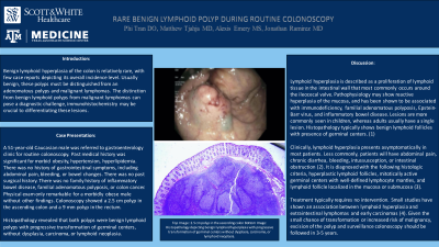
Case Description/Methods: A 51-year-old Caucasian male was referred to gastroenterology clinic for routine colonoscopy. Past medical history was significant for morbid obesity, hypertension, hyperlipidemia. There was no history of gastrointestinal symptoms, including abdominal pain, bleeding, or bowel changes. There was no past surgical history. There was no family history of inflammatory bowel disease, familial adenomatous polyposis, or colon cancer. Physical exam only remarkable for a morbidly obese male without other findings. Colonoscopy showed a 2.5 cm polyp in the ascending colon and a 9 mm polyp in the rectum.
Histopathology revealed that both polyps were benign lymphoid polyps with progressive transformation of germinal centers, without dysplasia, carcinoma, or lymphoid neoplasia.
Discussion: Lymphoid hyperplasia is described as a proliferation of lymphoid tissue in the intestinal wall that most commonly occurs around the ileocecal valve. Pathophysiology may show reactive hyperplasia of the mucosa, and has been shown to be associated with immunodeficiency, familial adenomatous polyposis, Epstein-Barr virus, and inflammatory bowel disease. Lesions are more commonly seen in children, whereas adults usually have a single lesion. Histopathology typically shows benign lymphoid follicles with presence of germinal centers. (1)
Clinically, lymphoid hyperplasia presents asymptomatically in most patients. Less commonly, patients will have abdominal pain, chronic diarrhea, bleeding, intussusception, or intestinal obstruction (2). It is diagnosed with the following histologic criteria, hyperplastic lymphoid follicles, mitotically active germinal centers with well-defined lymphocyte mantles, and lymphoid follicle localized in the mucosa or submucosa (3).
Treatment typically requires no intervention. Small studies have shown an association between lymphoid hyperplasia and extraintestinal lymphomas and early carcinomas (4). Given the small chance of transformation or increased risk of malignancy, excision of the polyp and surveillance colonoscopy should be followed in 3-5 years.

Disclosures:
Phi P. Tran, DO, Matthew Tjahja, MD, MS, Alexis Emery, BS, Jonathan Ramirez, MD. P0572 - Rare Benign Lymphoid Polyp During Routine Colonoscopy, ACG 2023 Annual Scientific Meeting Abstracts. Vancouver, BC, Canada: American College of Gastroenterology.
Baylor Scott & White, Temple, TX
Introduction: Benign lymphoid hyperplasia of the colon is relatively rare, with few case reports depicting its overall incidence level. Usually benign, these polyps must be distinguished from an adenomatous polyps and malignant lymphomas. The distinction from benign lymphoid polyps from malignant lymphomas can pose a diagnostic challenge, immunohistochemistry may be crucial to differentiating these lesions.
Case Description/Methods: A 51-year-old Caucasian male was referred to gastroenterology clinic for routine colonoscopy. Past medical history was significant for morbid obesity, hypertension, hyperlipidemia. There was no history of gastrointestinal symptoms, including abdominal pain, bleeding, or bowel changes. There was no past surgical history. There was no family history of inflammatory bowel disease, familial adenomatous polyposis, or colon cancer. Physical exam only remarkable for a morbidly obese male without other findings. Colonoscopy showed a 2.5 cm polyp in the ascending colon and a 9 mm polyp in the rectum.
Histopathology revealed that both polyps were benign lymphoid polyps with progressive transformation of germinal centers, without dysplasia, carcinoma, or lymphoid neoplasia.
Discussion: Lymphoid hyperplasia is described as a proliferation of lymphoid tissue in the intestinal wall that most commonly occurs around the ileocecal valve. Pathophysiology may show reactive hyperplasia of the mucosa, and has been shown to be associated with immunodeficiency, familial adenomatous polyposis, Epstein-Barr virus, and inflammatory bowel disease. Lesions are more commonly seen in children, whereas adults usually have a single lesion. Histopathology typically shows benign lymphoid follicles with presence of germinal centers. (1)
Clinically, lymphoid hyperplasia presents asymptomatically in most patients. Less commonly, patients will have abdominal pain, chronic diarrhea, bleeding, intussusception, or intestinal obstruction (2). It is diagnosed with the following histologic criteria, hyperplastic lymphoid follicles, mitotically active germinal centers with well-defined lymphocyte mantles, and lymphoid follicle localized in the mucosa or submucosa (3).
Treatment typically requires no intervention. Small studies have shown an association between lymphoid hyperplasia and extraintestinal lymphomas and early carcinomas (4). Given the small chance of transformation or increased risk of malignancy, excision of the polyp and surveillance colonoscopy should be followed in 3-5 years.

Figure: Top Image: 2.5 cm polyp in the ascending colon
Bottom Image: Histopathology depicting benign lymphoid hyperplasia with progressive transformation of germinal centers without dysplasia, carcinoma, or lymphoid neoplasia.
Bottom Image: Histopathology depicting benign lymphoid hyperplasia with progressive transformation of germinal centers without dysplasia, carcinoma, or lymphoid neoplasia.
Disclosures:
Phi Tran indicated no relevant financial relationships.
Matthew Tjahja indicated no relevant financial relationships.
Alexis Emery indicated no relevant financial relationships.
Jonathan Ramirez indicated no relevant financial relationships.
Phi P. Tran, DO, Matthew Tjahja, MD, MS, Alexis Emery, BS, Jonathan Ramirez, MD. P0572 - Rare Benign Lymphoid Polyp During Routine Colonoscopy, ACG 2023 Annual Scientific Meeting Abstracts. Vancouver, BC, Canada: American College of Gastroenterology.
