Sunday Poster Session
Category: General Endoscopy
P0585 - A Case of Pseudomelanosis Duodeni: Incidental Finding or Related to GI Bleed?
Sunday, October 22, 2023
3:30 PM - 7:00 PM PT
Location: Exhibit Hall

Has Audio

Afsha Aurshina, MD
UHS Wilson Hospital
Johnson City, NY
Presenting Author(s)
Afsha Aurshina, MD1, Vikram Ram Rajagopalan, MBBS2, Ibrar Atiq, MD3, Maria Bajwa, MD3, Minhaz Ahmad, MD3, Ali Marhaba, MD3
1UHS Wilson Hospital, Johnson City, NY; 2Kasturba Medical College, Manipal, Karnataka, India; 3United Health Services Hospital, Johnson City, NY
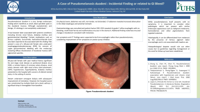
Introduction: Pseudomelanosis duodeni is a rare, benign endoscopic finding which manifests as small, black speckled spots in the duodenal mucosa. Although asymptomatic and benign, it’s etiology is not completely understood. It has however been associated with systemic conditions including chronic renal failure, diabetes mellitus and gastrointestinal bleeding. Certain medications such as ferrous sulphate, furosemide, hydrochlorothiazide have also been reported to be associated with this finding. We present a case of a 68-year-old female who underwent esophagogastroduodenoscopy (EGD) for concern of upper gastrointestinal bleeding with the endoscopy revealing diffuse discoloration of duodenal mucosa with pigmented speckles.
Case Description/Methods: 68-year-old female with past medical history significant for end stage renal disease on peritoneal dialysis since 2020, a remote history of coronary artery disease, renal artery stenosis with right renal artery stent placement, COPD, hypertension, hyperlipidemia, diabetes mellitus presented to the hospital with concern of altered mental status in the setting of uremia. Patient underwent emergent dialysis with subsequent improvement of mentation. However, her hospital course was complicated by multiple black tarry stools without a significant drop in hemoglobin from baseline ( 8 gm/dl). Abdomen was soft, non tender, not distended. CT abdomen revealed increased attenuation in the distal esophagus and proximal stomach. Gastroenterology was consulted and patient had an EGD. EGD revealed LA grade C reflux esophagitis with no bleeding, small hiatal hernia and erythematous mucosa in the stomach. Additional finding was mucosal changes in duodenum consistent with melanosis. Her symptoms and CT findings were suspected to be from esophagitis rather than pseudomelanosis coli considering improvement of her symptoms on proton pump inhibitor.
Discussion: While pseudomelanosis itself presents with no symptoms, it is important to consider other pathologies which can present with similar endoscopic appearance such as malignant melanoma, hemosiderosis and other pigmentations from ingested materials. Histologically, it can be differentiated from melanosis by the presence of ferrous pigment laden macrophages and is not associated with laxative use. Histopathological biopsies would rule out other causes but no guidelines regarding management or the need for follow up have been established.

Disclosures:
Afsha Aurshina, MD1, Vikram Ram Rajagopalan, MBBS2, Ibrar Atiq, MD3, Maria Bajwa, MD3, Minhaz Ahmad, MD3, Ali Marhaba, MD3. P0585 - A Case of Pseudomelanosis Duodeni: Incidental Finding or Related to GI Bleed?, ACG 2023 Annual Scientific Meeting Abstracts. Vancouver, BC, Canada: American College of Gastroenterology.
1UHS Wilson Hospital, Johnson City, NY; 2Kasturba Medical College, Manipal, Karnataka, India; 3United Health Services Hospital, Johnson City, NY
Introduction: Pseudomelanosis duodeni is a rare, benign endoscopic finding which manifests as small, black speckled spots in the duodenal mucosa. Although asymptomatic and benign, it’s etiology is not completely understood. It has however been associated with systemic conditions including chronic renal failure, diabetes mellitus and gastrointestinal bleeding. Certain medications such as ferrous sulphate, furosemide, hydrochlorothiazide have also been reported to be associated with this finding. We present a case of a 68-year-old female who underwent esophagogastroduodenoscopy (EGD) for concern of upper gastrointestinal bleeding with the endoscopy revealing diffuse discoloration of duodenal mucosa with pigmented speckles.
Case Description/Methods: 68-year-old female with past medical history significant for end stage renal disease on peritoneal dialysis since 2020, a remote history of coronary artery disease, renal artery stenosis with right renal artery stent placement, COPD, hypertension, hyperlipidemia, diabetes mellitus presented to the hospital with concern of altered mental status in the setting of uremia. Patient underwent emergent dialysis with subsequent improvement of mentation. However, her hospital course was complicated by multiple black tarry stools without a significant drop in hemoglobin from baseline ( 8 gm/dl). Abdomen was soft, non tender, not distended. CT abdomen revealed increased attenuation in the distal esophagus and proximal stomach. Gastroenterology was consulted and patient had an EGD. EGD revealed LA grade C reflux esophagitis with no bleeding, small hiatal hernia and erythematous mucosa in the stomach. Additional finding was mucosal changes in duodenum consistent with melanosis. Her symptoms and CT findings were suspected to be from esophagitis rather than pseudomelanosis coli considering improvement of her symptoms on proton pump inhibitor.
Discussion: While pseudomelanosis itself presents with no symptoms, it is important to consider other pathologies which can present with similar endoscopic appearance such as malignant melanoma, hemosiderosis and other pigmentations from ingested materials. Histologically, it can be differentiated from melanosis by the presence of ferrous pigment laden macrophages and is not associated with laxative use. Histopathological biopsies would rule out other causes but no guidelines regarding management or the need for follow up have been established.

Figure: Endoscopic imaging showing speckled pigmentation pattern in duodenum
Disclosures:
Afsha Aurshina indicated no relevant financial relationships.
Vikram Ram Rajagopalan indicated no relevant financial relationships.
Ibrar Atiq indicated no relevant financial relationships.
Maria Bajwa indicated no relevant financial relationships.
Minhaz Ahmad indicated no relevant financial relationships.
Ali Marhaba indicated no relevant financial relationships.
Afsha Aurshina, MD1, Vikram Ram Rajagopalan, MBBS2, Ibrar Atiq, MD3, Maria Bajwa, MD3, Minhaz Ahmad, MD3, Ali Marhaba, MD3. P0585 - A Case of Pseudomelanosis Duodeni: Incidental Finding or Related to GI Bleed?, ACG 2023 Annual Scientific Meeting Abstracts. Vancouver, BC, Canada: American College of Gastroenterology.
