Sunday Poster Session
Category: GI Bleeding
P0623 - Management of Life Threatening GI Bleeding Due to Cameron Lesions: A Case Report
Sunday, October 22, 2023
3:30 PM - 7:00 PM PT
Location: Exhibit Hall

Has Audio

Yazan Abboud, MD
Rutgers New Jersey Medical School
Newark, NJ
Presenting Author(s)
Ahmed Abdulrahman, MD1, Tarek Aboursheid, MD1, Monzer Abdalla, MD1, Yazan Abboud, MD2
1Ascension Saint Francis Hospital, Evanston, IL; 2Rutgers New Jersey Medical School, Newark, NJ
Introduction: Cameron lesions are mucosal erosions and ulcerations that develop in individuals with large hiatal hernias. They are often considered incidental findings during endoscopy or can lead to chronic GI bleeding, but acute bleeding is uncommon. We present a case of acute, life-threatening GI bleeding due to Cameron lesions that were managed successfully.
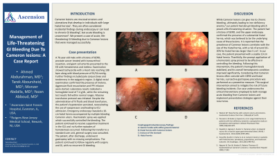
Case Description/Methods: A 76-year-old male with a history of GERD, prostate cancer treated with transurethral resection, and giant cell arteritis presented to the ED with hematemesis and melena. Examination showed tachycardia with HR of 108, blood pressure of 91/54 mmHg, pale conjunctivae, tenderness in the epigastric region, and digital rectal examination revealed melena. Aggressive fluid resuscitation and PPI infusion were initiated. Lab results showed an hgb level of 7.4 g/dL but otherwise were normal. Massive transfusion protocol was initiated, but the patient remained hypotensive and required vasopressors, leading to ICU admission. Emergency endoscopy revealed a 10-cm hiatal hernia with multiple bleeding Cameron ulcers. Hemostatic spray was applied which successfully controlled the bleeding. The patient received continued supportive treatment in the ICU, without further hematemesis episodes. After transfer to the floor, general surgery was consulted and the patient underwent gastropexy without complications, followed by regular follow-up with surgery and GI, with no recurrent GI bleeding.
Discussion: Cameron lesions, although typically an incidental finding in patients with hiatal hernia, can lead to chronic bleeding and result in iron deficiency anemia. In our patient, the bleeding caused by Cameron lesions was acute and life-threatening. The patient had a history of GERD, and the endoscopy confirmed the presence of a large hiatal hernia, which was believed to be the underlying cause of these lesions. It is reported that the prevalence of Cameron lesions correlates with the size of the hiatal hernia, with a risk of 10–20% for hiatal hernias larger than 5 cm. In our case, the patient had a 10 cm hiatal hernia. Thankfully, the bleeding was effectively controlled using a hemostatic spray. after which the patient’s hemoglobin remained stable and his hemodynamic state improved. Considering that Cameron lesions are commonly associated with reflux esophagitis and hiatal hernia a gastropexy procedure was performed as a preventive treatment. Our case highlights important measures to effectively treat and prevent further bleeding from Cameron lesions.

Disclosures:
Ahmed Abdulrahman, MD1, Tarek Aboursheid, MD1, Monzer Abdalla, MD1, Yazan Abboud, MD2. P0623 - Management of Life Threatening GI Bleeding Due to Cameron Lesions: A Case Report, ACG 2023 Annual Scientific Meeting Abstracts. Vancouver, BC, Canada: American College of Gastroenterology.
1Ascension Saint Francis Hospital, Evanston, IL; 2Rutgers New Jersey Medical School, Newark, NJ
Introduction: Cameron lesions are mucosal erosions and ulcerations that develop in individuals with large hiatal hernias. They are often considered incidental findings during endoscopy or can lead to chronic GI bleeding, but acute bleeding is uncommon. We present a case of acute, life-threatening GI bleeding due to Cameron lesions that were managed successfully.
Case Description/Methods: A 76-year-old male with a history of GERD, prostate cancer treated with transurethral resection, and giant cell arteritis presented to the ED with hematemesis and melena. Examination showed tachycardia with HR of 108, blood pressure of 91/54 mmHg, pale conjunctivae, tenderness in the epigastric region, and digital rectal examination revealed melena. Aggressive fluid resuscitation and PPI infusion were initiated. Lab results showed an hgb level of 7.4 g/dL but otherwise were normal. Massive transfusion protocol was initiated, but the patient remained hypotensive and required vasopressors, leading to ICU admission. Emergency endoscopy revealed a 10-cm hiatal hernia with multiple bleeding Cameron ulcers. Hemostatic spray was applied which successfully controlled the bleeding. The patient received continued supportive treatment in the ICU, without further hematemesis episodes. After transfer to the floor, general surgery was consulted and the patient underwent gastropexy without complications, followed by regular follow-up with surgery and GI, with no recurrent GI bleeding.
Discussion: Cameron lesions, although typically an incidental finding in patients with hiatal hernia, can lead to chronic bleeding and result in iron deficiency anemia. In our patient, the bleeding caused by Cameron lesions was acute and life-threatening. The patient had a history of GERD, and the endoscopy confirmed the presence of a large hiatal hernia, which was believed to be the underlying cause of these lesions. It is reported that the prevalence of Cameron lesions correlates with the size of the hiatal hernia, with a risk of 10–20% for hiatal hernias larger than 5 cm. In our case, the patient had a 10 cm hiatal hernia. Thankfully, the bleeding was effectively controlled using a hemostatic spray. after which the patient’s hemoglobin remained stable and his hemodynamic state improved. Considering that Cameron lesions are commonly associated with reflux esophagitis and hiatal hernia a gastropexy procedure was performed as a preventive treatment. Our case highlights important measures to effectively treat and prevent further bleeding from Cameron lesions.

Figure: Image A: shows the Gastric Cardia. Images B&C: show Cameron lesions, and Image D: shows Hiatal Hernia
Disclosures:
Ahmed Abdulrahman indicated no relevant financial relationships.
Tarek Aboursheid indicated no relevant financial relationships.
Monzer Abdalla indicated no relevant financial relationships.
Yazan Abboud indicated no relevant financial relationships.
Ahmed Abdulrahman, MD1, Tarek Aboursheid, MD1, Monzer Abdalla, MD1, Yazan Abboud, MD2. P0623 - Management of Life Threatening GI Bleeding Due to Cameron Lesions: A Case Report, ACG 2023 Annual Scientific Meeting Abstracts. Vancouver, BC, Canada: American College of Gastroenterology.
