Sunday Poster Session
Category: GI Bleeding
P0643 - Arterio-Venous Malformation Masquerading as Small Bowel Mass: A Rare Presentation of Overt GI Bleeding
Sunday, October 22, 2023
3:30 PM - 7:00 PM PT
Location: Exhibit Hall

Has Audio
- KO
Kashif R. Osmani, MD
University of Illinois at Chicago
Chicago, Illinois
Presenting Author(s)
Kashif R. Osmani, MD1, Sana Hussain, MD2, Snehal Sonawane, MD1, Michael Gianarakis, MD1, Saeed Ali, MD3, Brian Boulay, MD, MPH1
1University of Illinois at Chicago, Chicago, IL; 2Khyber Teaching Hospital, Peshawar, Northern Areas, Pakistan; 3University of Illinois Chicago, Chicago, IL
Introduction: Obscure gastrointestinal bleeding (OGIB) refers to GI bleeding of unknown origin despite thorough evaluation of upper and lower GI tract. It constitutes 5% of all GI bleeds. It includes overt bleeding (melena or hematochezia) and occult bleeding. The diagnosis of small bowel bleeding from arterio-venous malformations (AVM) poses significant difficulty, and diagnosis and management can be challenging. We present a rare case of overt GI bleeding from an AVM masquerading as a small bowel mass.
Case Description/Methods: 76-year-old female with history of end stage renal disease on hemodialysis, hypertension, type-2 diabetes, and hyperlipidemia presented with a 3-week history of fatigue, black stools, and abdominal pain. Medications included Aspirin 81 mg daily. Rectal exam showed melenic stools. Lab results notable for Hgb 5.7 (baseline ~10) with MCV of 100, platelets 174, BUN 27, Cr 3.31, and Lactate 1.2. Patient received blood transfusion and Protonix drip. Colonoscopy done a year ago showed diverticulosis and two small polyps (tubular adenomas). Upper GI endoscopy performed on admission was unremarkable. Video capsule endoscopy revealed a medium-sized 1-cm mass in the jejunum with ulceration and signs of recent bleeding. An antegrade double-balloon enteroscopy showed a 13-mm submucosal and ulcerated jejunal mass. Biopsy attempts triggered bleeding requiring hemostatic clips. CT abdomen pelvis confirmed jejunal mass adjacent to the clips. Patient subsequently required exploratory laparotomy, a palpable jejunal mass was felt, and segmental jejunal resection was performed. Pathology of the resected small intestine and jejunum showed consistency with arteriovenous malformation along with mucosal ulceration, hemorrhage, and granulation tissue.
Discussion: Bleeding from small bowel AVMs can be life threatening, requiring surgical resection at times due to risk of re-bleeding. Per the Yano-Yamamoto classification of small bowel vascular lesions, Type 1 AVMs present as punctate pulsatile lesions, often in elderly patients, and is an acquired disease. On the contrary, Type 4 AVMs occur in younger patients with congenital intestinal AVMs and can be mass-like. Our case, however, shows a rare presentation of an AVM presenting as mass like AVM with mucosal ulceration, needing resection for high suspicion of tumor with risk of re-bleeding. This demonstrates the diligence needed for thorough evaluation of atypical AVM’s due to the high risk of future complications.

Disclosures:
Kashif R. Osmani, MD1, Sana Hussain, MD2, Snehal Sonawane, MD1, Michael Gianarakis, MD1, Saeed Ali, MD3, Brian Boulay, MD, MPH1. P0643 - Arterio-Venous Malformation Masquerading as Small Bowel Mass: A Rare Presentation of Overt GI Bleeding, ACG 2023 Annual Scientific Meeting Abstracts. Vancouver, BC, Canada: American College of Gastroenterology.
1University of Illinois at Chicago, Chicago, IL; 2Khyber Teaching Hospital, Peshawar, Northern Areas, Pakistan; 3University of Illinois Chicago, Chicago, IL
Introduction: Obscure gastrointestinal bleeding (OGIB) refers to GI bleeding of unknown origin despite thorough evaluation of upper and lower GI tract. It constitutes 5% of all GI bleeds. It includes overt bleeding (melena or hematochezia) and occult bleeding. The diagnosis of small bowel bleeding from arterio-venous malformations (AVM) poses significant difficulty, and diagnosis and management can be challenging. We present a rare case of overt GI bleeding from an AVM masquerading as a small bowel mass.
Case Description/Methods: 76-year-old female with history of end stage renal disease on hemodialysis, hypertension, type-2 diabetes, and hyperlipidemia presented with a 3-week history of fatigue, black stools, and abdominal pain. Medications included Aspirin 81 mg daily. Rectal exam showed melenic stools. Lab results notable for Hgb 5.7 (baseline ~10) with MCV of 100, platelets 174, BUN 27, Cr 3.31, and Lactate 1.2. Patient received blood transfusion and Protonix drip. Colonoscopy done a year ago showed diverticulosis and two small polyps (tubular adenomas). Upper GI endoscopy performed on admission was unremarkable. Video capsule endoscopy revealed a medium-sized 1-cm mass in the jejunum with ulceration and signs of recent bleeding. An antegrade double-balloon enteroscopy showed a 13-mm submucosal and ulcerated jejunal mass. Biopsy attempts triggered bleeding requiring hemostatic clips. CT abdomen pelvis confirmed jejunal mass adjacent to the clips. Patient subsequently required exploratory laparotomy, a palpable jejunal mass was felt, and segmental jejunal resection was performed. Pathology of the resected small intestine and jejunum showed consistency with arteriovenous malformation along with mucosal ulceration, hemorrhage, and granulation tissue.
Discussion: Bleeding from small bowel AVMs can be life threatening, requiring surgical resection at times due to risk of re-bleeding. Per the Yano-Yamamoto classification of small bowel vascular lesions, Type 1 AVMs present as punctate pulsatile lesions, often in elderly patients, and is an acquired disease. On the contrary, Type 4 AVMs occur in younger patients with congenital intestinal AVMs and can be mass-like. Our case, however, shows a rare presentation of an AVM presenting as mass like AVM with mucosal ulceration, needing resection for high suspicion of tumor with risk of re-bleeding. This demonstrates the diligence needed for thorough evaluation of atypical AVM’s due to the high risk of future complications.

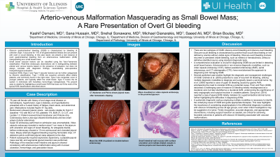
Figure: Image A: CT Abdomen and Pelvis shows jejunal mass after hemostasis clipping
Image B: Mass visualized on video capsule endoscopy in the proximal jejunum
Image C: Mass-like AVM visualized on double-balloon enteroscopy
Image B: Mass visualized on video capsule endoscopy in the proximal jejunum
Image C: Mass-like AVM visualized on double-balloon enteroscopy
Disclosures:
Kashif Osmani indicated no relevant financial relationships.
Sana Hussain indicated no relevant financial relationships.
Snehal Sonawane indicated no relevant financial relationships.
Michael Gianarakis indicated no relevant financial relationships.
Saeed Ali indicated no relevant financial relationships.
Brian Boulay indicated no relevant financial relationships.
Kashif R. Osmani, MD1, Sana Hussain, MD2, Snehal Sonawane, MD1, Michael Gianarakis, MD1, Saeed Ali, MD3, Brian Boulay, MD, MPH1. P0643 - Arterio-Venous Malformation Masquerading as Small Bowel Mass: A Rare Presentation of Overt GI Bleeding, ACG 2023 Annual Scientific Meeting Abstracts. Vancouver, BC, Canada: American College of Gastroenterology.

