Sunday Poster Session
Category: GI Bleeding
P0670 - A Unique Presentation of CMV Colitis in Acute GI Bleed
Sunday, October 22, 2023
3:30 PM - 7:00 PM PT
Location: Exhibit Hall

Has Audio

Dhruv Patel, DO
St. Joseph's University Medical Center
Paterson, New Jersey
Presenting Author(s)
Dhruv Patel, DO1, Dawid Nowak, MD2, Nida Ansari, DO2, Angela Pais Vidreiro, BS, 3, Gabriel Melki, MD2, Yana Cavanagh, MD1
1St. Joseph's University Medical Center, Paterson, NJ; 2St. Joseph's University Medical Center, Paterson, NJ; 3St. Joseph’s University Medical Center, Paterson, NJ
Introduction: CMV colitis is a relatively common end-organ infectious complication in immunocompromised hosts which negatively affects clinical outcomes. Presentation typically includes abdominal pain and bloody diarrhea. Diagnosis methods include tissue biopsy, culture testing, and PCR. Immunohistochemistry is the most reliable method and can identify "owl eye appearance" inclusion bodies. Endoscopic exams may reveal ulcerations, mucous edema, and inflamed mucosa. Patients over 55 with underlying conditions have a higher mortality risk and early treatment can lead to decreased morbidity and mortality in CMV colitis.
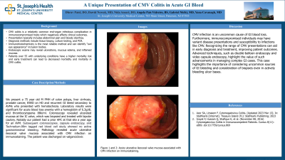
Case Description/Methods: We present a 75 year old M PMH of colon polyps, liver cirrhosis, prostate cancer, ESRD on HD and recurrent GI bleed secondary to AVMs who presented with hematochezia. Laboratory results were significant for acute blood loss anemia with a hemoglobin of 6.3g/dL and thrombocytopenia 88k/ml. Colonoscopy revealed ulcerated mucosa at the IC valve, which was biopsied and treated with bipolar cautery. Notably our patient had a prior APC at that site a year ago for an AVM. Pathology revealed acute ulcerative ileocecal valve mucosa associated with CMV infection on immunostaining. The patient was discharged on valganciclovir.
Discussion: CMV infection is an uncommon cause of GI blood loss. Furthermore, immunocompromised individuals may have variant disease presentations and susceptibility to infections like CMV. Recognizing the range of CMV presentations can aid in early diagnosis and treatment, improving patient outcomes. This case highlights the importance of considering uncommon sources of GI bleeding and consideration of biopsies even in actively bleeding ulcer bases.

Disclosures:
Dhruv Patel, DO1, Dawid Nowak, MD2, Nida Ansari, DO2, Angela Pais Vidreiro, BS, 3, Gabriel Melki, MD2, Yana Cavanagh, MD1. P0670 - A Unique Presentation of CMV Colitis in Acute GI Bleed, ACG 2023 Annual Scientific Meeting Abstracts. Vancouver, BC, Canada: American College of Gastroenterology.
1St. Joseph's University Medical Center, Paterson, NJ; 2St. Joseph's University Medical Center, Paterson, NJ; 3St. Joseph’s University Medical Center, Paterson, NJ
Introduction: CMV colitis is a relatively common end-organ infectious complication in immunocompromised hosts which negatively affects clinical outcomes. Presentation typically includes abdominal pain and bloody diarrhea. Diagnosis methods include tissue biopsy, culture testing, and PCR. Immunohistochemistry is the most reliable method and can identify "owl eye appearance" inclusion bodies. Endoscopic exams may reveal ulcerations, mucous edema, and inflamed mucosa. Patients over 55 with underlying conditions have a higher mortality risk and early treatment can lead to decreased morbidity and mortality in CMV colitis.
Case Description/Methods: We present a 75 year old M PMH of colon polyps, liver cirrhosis, prostate cancer, ESRD on HD and recurrent GI bleed secondary to AVMs who presented with hematochezia. Laboratory results were significant for acute blood loss anemia with a hemoglobin of 6.3g/dL and thrombocytopenia 88k/ml. Colonoscopy revealed ulcerated mucosa at the IC valve, which was biopsied and treated with bipolar cautery. Notably our patient had a prior APC at that site a year ago for an AVM. Pathology revealed acute ulcerative ileocecal valve mucosa associated with CMV infection on immunostaining. The patient was discharged on valganciclovir.
Discussion: CMV infection is an uncommon cause of GI blood loss. Furthermore, immunocompromised individuals may have variant disease presentations and susceptibility to infections like CMV. Recognizing the range of CMV presentations can aid in early diagnosis and treatment, improving patient outcomes. This case highlights the importance of considering uncommon sources of GI bleeding and consideration of biopsies even in actively bleeding ulcer bases.

Figure: Acute ulcerative ileocecal valve mucosa associated with CMV infection on immunostaining.
Disclosures:
Dhruv Patel indicated no relevant financial relationships.
Dawid Nowak indicated no relevant financial relationships.
Nida Ansari indicated no relevant financial relationships.
Angela Pais Vidreiro, BS indicated no relevant financial relationships.
Gabriel Melki indicated no relevant financial relationships.
Yana Cavanagh indicated no relevant financial relationships.
Dhruv Patel, DO1, Dawid Nowak, MD2, Nida Ansari, DO2, Angela Pais Vidreiro, BS, 3, Gabriel Melki, MD2, Yana Cavanagh, MD1. P0670 - A Unique Presentation of CMV Colitis in Acute GI Bleed, ACG 2023 Annual Scientific Meeting Abstracts. Vancouver, BC, Canada: American College of Gastroenterology.
