Sunday Poster Session
Category: IBD
P0801 - Toxic Positivity: An Incidental Discovery of Crohn’s Disease in Acute Abdominal Distention
Sunday, October 22, 2023
3:30 PM - 7:00 PM PT
Location: Exhibit Hall

Has Audio

Jeff Taclob, MD
Texas Tech University Health Sciences Center
El Paso, TX
Presenting Author(s)
Jeff Angelo Taclob, MD, Gian Marco Galura, MD, Brian Lee, DO, Bhavi Trivedi, MD, Nawar Hakim, MD, Marc Zuckerman, MD, Ihsan Al Bayati, MD
Texas Tech University Health Sciences Center, El Paso, TX
Introduction: Toxic Megacolon (TM) is a rare but potentially fatal complication of colonic inflammation, defined as acute colonic dilatation of >6cm with accompanying systemic toxicity, inflammation, or infectious condition. Frequency increases with age and is most commonly associated with Inflammatory Bowel Disease (IBD), septicemia, and intestinal factors. Crohn’s disease (CD) is a chronic condition characterized by transmural inflammation that affects regions of the gastrointestinal tract, involving any part from the mouth down to the anus with the most common regions affected being the small intestine and distal ileum. This report shows the correlation caused by an underlying inflammatory condition leading to the development of toxic megacolon.
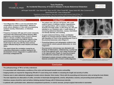
Case Description/Methods: The patient was a 56-year-old female, with type 2 diabetes mellitus, hypertension, and hypothyroidism. She denied smoking, alcoholic beverage intake, or illicit drug use. She was admitted to our institution with a 1-week history of worsening abdominal distention, nausea, intermittent non-bloody diarrhea, and vomiting. A supine abdominal Kidney, Ureter, and Bladder (KUB) X-ray showed extensive dilation of the bowel, with diameters of 13cm in the ascending and 11cm in the proximal transverse colon (Figure 1). Features were concerning for TM vs Colonic Pseudo-obstruction. Decompressive colonoscopy was attempted with findings of inflammation, cobblestoning, a partial tear in the recto-sigmoid colon, and a deep mucosal tear in the sigmoid colon requiring emergent total abdominal colectomy with end ileostomy. Histological analysis showed multiple areas with features consistent with Crohn’s colitis.
Discussion: The pathophysiology of TM is not fully understood. It is associated with inflammatory conditions of the colon and decreased smooth muscle contractility. Imaging studies are required for diagnosing TM with CT scans being more reliable in evaluating the length and severity of colitis. Plain abdominal radiograph features include colonic diameter >6cm (rarely >15cm) with the ascending and transverse colon as being the most dilated. The main objectives are attenuation of colitis, correction of electrolyte derangements, anemia, dehydration, toxemia, and preventing bowel perforation. Infectious causes should be ruled out before initiating standard therapy with IV (Intravenous) steroids. In patients with a perforated colon, abdominal compartment syndrome, or colonic necrosis, prompt surgical intervention is warranted.

Disclosures:
Jeff Angelo Taclob, MD, Gian Marco Galura, MD, Brian Lee, DO, Bhavi Trivedi, MD, Nawar Hakim, MD, Marc Zuckerman, MD, Ihsan Al Bayati, MD. P0801 - Toxic Positivity: An Incidental Discovery of Crohn’s Disease in Acute Abdominal Distention, ACG 2023 Annual Scientific Meeting Abstracts. Vancouver, BC, Canada: American College of Gastroenterology.
Texas Tech University Health Sciences Center, El Paso, TX
Introduction: Toxic Megacolon (TM) is a rare but potentially fatal complication of colonic inflammation, defined as acute colonic dilatation of >6cm with accompanying systemic toxicity, inflammation, or infectious condition. Frequency increases with age and is most commonly associated with Inflammatory Bowel Disease (IBD), septicemia, and intestinal factors. Crohn’s disease (CD) is a chronic condition characterized by transmural inflammation that affects regions of the gastrointestinal tract, involving any part from the mouth down to the anus with the most common regions affected being the small intestine and distal ileum. This report shows the correlation caused by an underlying inflammatory condition leading to the development of toxic megacolon.
Case Description/Methods: The patient was a 56-year-old female, with type 2 diabetes mellitus, hypertension, and hypothyroidism. She denied smoking, alcoholic beverage intake, or illicit drug use. She was admitted to our institution with a 1-week history of worsening abdominal distention, nausea, intermittent non-bloody diarrhea, and vomiting. A supine abdominal Kidney, Ureter, and Bladder (KUB) X-ray showed extensive dilation of the bowel, with diameters of 13cm in the ascending and 11cm in the proximal transverse colon (Figure 1). Features were concerning for TM vs Colonic Pseudo-obstruction. Decompressive colonoscopy was attempted with findings of inflammation, cobblestoning, a partial tear in the recto-sigmoid colon, and a deep mucosal tear in the sigmoid colon requiring emergent total abdominal colectomy with end ileostomy. Histological analysis showed multiple areas with features consistent with Crohn’s colitis.
Discussion: The pathophysiology of TM is not fully understood. It is associated with inflammatory conditions of the colon and decreased smooth muscle contractility. Imaging studies are required for diagnosing TM with CT scans being more reliable in evaluating the length and severity of colitis. Plain abdominal radiograph features include colonic diameter >6cm (rarely >15cm) with the ascending and transverse colon as being the most dilated. The main objectives are attenuation of colitis, correction of electrolyte derangements, anemia, dehydration, toxemia, and preventing bowel perforation. Infectious causes should be ruled out before initiating standard therapy with IV (Intravenous) steroids. In patients with a perforated colon, abdominal compartment syndrome, or colonic necrosis, prompt surgical intervention is warranted.

Figure: Supine Kidney, Ureter, and Bladder X-ray. Extensive dilation of the colon measuring up to 13 cm in the ascending colon and 11 cm in the proximal transverse colon with no free air identified
Disclosures:
Jeff Angelo Taclob indicated no relevant financial relationships.
Gian Marco Galura indicated no relevant financial relationships.
Brian Lee indicated no relevant financial relationships.
Bhavi Trivedi indicated no relevant financial relationships.
Nawar Hakim indicated no relevant financial relationships.
Marc Zuckerman indicated no relevant financial relationships.
Ihsan Al Bayati indicated no relevant financial relationships.
Jeff Angelo Taclob, MD, Gian Marco Galura, MD, Brian Lee, DO, Bhavi Trivedi, MD, Nawar Hakim, MD, Marc Zuckerman, MD, Ihsan Al Bayati, MD. P0801 - Toxic Positivity: An Incidental Discovery of Crohn’s Disease in Acute Abdominal Distention, ACG 2023 Annual Scientific Meeting Abstracts. Vancouver, BC, Canada: American College of Gastroenterology.
