Sunday Poster Session
Category: IBD
P0803 - Yersiniosis: An Overlooked Mimicker of Crohn's Disease
Sunday, October 22, 2023
3:30 PM - 7:00 PM PT
Location: Exhibit Hall

Has Audio

Siri Urquhart, MD
Mayo Clinic
Rochester, MN
Presenting Author(s)
Shubham Sood, MBBS, Siri Urquhart, MD, Nayantara Coelho-Prabhu, MBBS
Mayo Clinic, Rochester, MN
Introduction: Crohn’s disease (CD) is a chronic, idiopathic immune-mediated condition involving the gastrointestinal (GI) tract which can present with a wide range of symptoms. A combination of clinical, laboratory, endoscopic, and radiographic findings are used to exclude other etiologies and confirm the diagnosis. The differential diagnosis of CD can be quite broad, consisting of both noninfectious and infectious mimics. However, the diagnosis of CD can be difficult to establish given its nonspecific clinical symptoms and varying endoscopic and histologic findings.
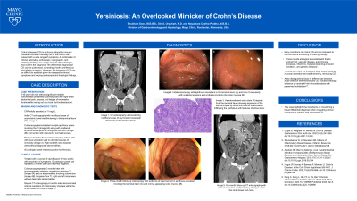
Case Description/Methods: A 45-year-old man presented to primary care with right-sided abdominal pain, nausea, and fatigue of two weeks duration after eating out at a local fast-food restaurant. CRP was found to be mildly elevated at 11 mg/L. CT enterography demonstrated multifocal areas of asymmetric bowel wall thickening in the terminal ileum (TI). Colonoscopy demonstrated multiple aphthous ulcers involving the TI (Figure 1A) along with scattered erosions and erythema throughout the colon and rectum with intervening normal mucosa (Figure 1B). Biopsies from the TI revealed moderately active ileitis with focal ulceration and no definite features of chronicity. Right and left colon biopsies were without diagnostic abnormalities. GI pathogen panel was then obtained which returned positive for Yersinia species. The patient was started on a course of ciprofloxacin for two weeks. Two months later, GI pathogen panel was repeated which was negative. Colonoscopy was repeated three months later which demonstrated improvement in aphthous ulcerations involving the TI (Figure 1C) and no abnormalities involving the colorectum (Figure 1D). Biopsies from the TI, right, and left colon were without diagnostic abnormalities.
Discussion: Many conditions can mimic CD and are important to rule out before embarking on a lifelong therapy. These include etiologies associated with the GI luminal tract, vascular disease, autoimmune processes, infections, malignancies, drug-induced conditions, and genetic diseases. Yersinia can infect the small and large bowel, causing mucosal ulceration and wall thickening, mimicking CD. However, a key distinguishing factor to differentiate between acute infection with Yersinia and CD includes histologic evidence of neutrophil-rich microabscesses with preserved architecture. This case highlights the importance of considering a broad differential diagnosis when evaluating clinical symptoms in patients with suspected CD.

Disclosures:
Shubham Sood, MBBS, Siri Urquhart, MD, Nayantara Coelho-Prabhu, MBBS. P0803 - Yersiniosis: An Overlooked Mimicker of Crohn's Disease, ACG 2023 Annual Scientific Meeting Abstracts. Vancouver, BC, Canada: American College of Gastroenterology.
Mayo Clinic, Rochester, MN
Introduction: Crohn’s disease (CD) is a chronic, idiopathic immune-mediated condition involving the gastrointestinal (GI) tract which can present with a wide range of symptoms. A combination of clinical, laboratory, endoscopic, and radiographic findings are used to exclude other etiologies and confirm the diagnosis. The differential diagnosis of CD can be quite broad, consisting of both noninfectious and infectious mimics. However, the diagnosis of CD can be difficult to establish given its nonspecific clinical symptoms and varying endoscopic and histologic findings.
Case Description/Methods: A 45-year-old man presented to primary care with right-sided abdominal pain, nausea, and fatigue of two weeks duration after eating out at a local fast-food restaurant. CRP was found to be mildly elevated at 11 mg/L. CT enterography demonstrated multifocal areas of asymmetric bowel wall thickening in the terminal ileum (TI). Colonoscopy demonstrated multiple aphthous ulcers involving the TI (Figure 1A) along with scattered erosions and erythema throughout the colon and rectum with intervening normal mucosa (Figure 1B). Biopsies from the TI revealed moderately active ileitis with focal ulceration and no definite features of chronicity. Right and left colon biopsies were without diagnostic abnormalities. GI pathogen panel was then obtained which returned positive for Yersinia species. The patient was started on a course of ciprofloxacin for two weeks. Two months later, GI pathogen panel was repeated which was negative. Colonoscopy was repeated three months later which demonstrated improvement in aphthous ulcerations involving the TI (Figure 1C) and no abnormalities involving the colorectum (Figure 1D). Biopsies from the TI, right, and left colon were without diagnostic abnormalities.
Discussion: Many conditions can mimic CD and are important to rule out before embarking on a lifelong therapy. These include etiologies associated with the GI luminal tract, vascular disease, autoimmune processes, infections, malignancies, drug-induced conditions, and genetic diseases. Yersinia can infect the small and large bowel, causing mucosal ulceration and wall thickening, mimicking CD. However, a key distinguishing factor to differentiate between acute infection with Yersinia and CD includes histologic evidence of neutrophil-rich microabscesses with preserved architecture. This case highlights the importance of considering a broad differential diagnosis when evaluating clinical symptoms in patients with suspected CD.

Figure: Figure 1. Initial colonoscopy with aphthous ulcerations in terminal ileum (A) and scattered loss of vascularity, erosions, and erythema involving the colon mucosa (B). Follow-up colonoscopy three months later with evidence of improvement in aphthous ulcerations involving the terminal ileum (C) and normal-appearing colon mucosa (D).
Disclosures:
Shubham Sood indicated no relevant financial relationships.
Siri Urquhart indicated no relevant financial relationships.
Nayantara Coelho-Prabhu: Alexion Pharma – Advisor or Review Panel Member. Iterative Health – Advisor or Review Panel Member.
Shubham Sood, MBBS, Siri Urquhart, MD, Nayantara Coelho-Prabhu, MBBS. P0803 - Yersiniosis: An Overlooked Mimicker of Crohn's Disease, ACG 2023 Annual Scientific Meeting Abstracts. Vancouver, BC, Canada: American College of Gastroenterology.
