Sunday Poster Session
Category: IBD
P0811 - Synchronous Low-Grade Tubulogalndular Adenocarcinoma and High-Grade Adenocarcinoma in a 28-Year-Old Male with Ulcerative Colitis
Sunday, October 22, 2023
3:30 PM - 7:00 PM PT
Location: Exhibit Hall

Has Audio

Kevin Turner, DO
University of Minnesota
Minneapolis, Minnesota
Presenting Author(s)
Mohammad A. Alutaibi, MBBS1, Khalid Amin, MD2, Oyedele Adeyi, MD3, Kevin Turner, DO1
1University of Minnesota, Minneapolis, MN; 2University of Minnesota Medical Center, Minneapolis, MN; 3University of Minnesota Medical School, Minneapolis, MN
Introduction: Inflammatory bowel disease (IBD) is a well-known risk factor for dysplasia and malignancy, especially in patients with young onset and extensive disease. One type of adenocarcinoma associated with IBD is low-grade tubulogalndular adenocarcinoma (LGTGA) which can arise directly from low-grade dysplasia and may transform into a higher-grade carcinoma. LGTGA is a neoplasm seen almost exclusively in IBD patients, accounting for 11% of all lower gastrointestinal tract carcinomas in these patients. It may present macroscopically as a stenotic, exophytic, or flat lesion.
Case Description/Methods: We present a 28-year-old male patient with a long-standing panulcerative colitis on Infliximab, whose disease continued to worsen despite switching to Vedolizumab, leading ultimately to total abdominal colectomy with end ileostomy due to uncontrolled disease. A prior surveillance colonoscopy had revealed severe colitis with no dysplasia or malignancy. There was no family history of IBD, but there was a family history of uterine cancer and multiple myeloma.
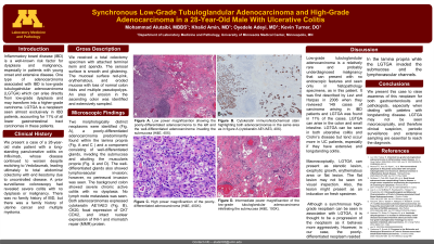
The colectomy specimen showed loss of normal colon folds and multiple pseudopolyps. An area of erosion in the ascending colon was identified and extensively sampled. Microscopically, two morphologically distinct neoplasms were identified (Fig. A), a poorly-differentiated adenocarcinoma predominantly within the lamina propria (Fig. A and C ) and a component consisting of well-differentiated glands, invading the submucosa and abutting the muscularis propria (Fig. A and D). The well-differentiated glands also showed lymphovascular invasion; however, no perineural invasion was seen. The background colon showed severe chronic active colitis with no dysplasia. Both adenocarcinomas expressed AE1/AE3 (Fig. B), CDX2, CK20, cytokeratin, focal expression of CK7, and intact nuclear expression of INI-1 and MMR.
Discussion: LGTGA is a relatively rare and probably underdiagnosed malignancy that can present with no endoscopic features and seen only in histopathology specimens, as in this patient. Although a high-grade neoplasm can be seen in association with LGTGA, it is thought to be a progression of the neoplasm as it behaves more aggressively; however, in our case, the poorly differentiated neoplasm resided in the lamina propria while the LGTGA invaded the submucosa and lymphovascular vessels. Therefore, for the fourth mentioned reasons, it is of importance to have a high level of suspicion in IBD biopsy and resection assessments.

Disclosures:
Mohammad A. Alutaibi, MBBS1, Khalid Amin, MD2, Oyedele Adeyi, MD3, Kevin Turner, DO1. P0811 - Synchronous Low-Grade Tubulogalndular Adenocarcinoma and High-Grade Adenocarcinoma in a 28-Year-Old Male with Ulcerative Colitis, ACG 2023 Annual Scientific Meeting Abstracts. Vancouver, BC, Canada: American College of Gastroenterology.
1University of Minnesota, Minneapolis, MN; 2University of Minnesota Medical Center, Minneapolis, MN; 3University of Minnesota Medical School, Minneapolis, MN
Introduction: Inflammatory bowel disease (IBD) is a well-known risk factor for dysplasia and malignancy, especially in patients with young onset and extensive disease. One type of adenocarcinoma associated with IBD is low-grade tubulogalndular adenocarcinoma (LGTGA) which can arise directly from low-grade dysplasia and may transform into a higher-grade carcinoma. LGTGA is a neoplasm seen almost exclusively in IBD patients, accounting for 11% of all lower gastrointestinal tract carcinomas in these patients. It may present macroscopically as a stenotic, exophytic, or flat lesion.
Case Description/Methods: We present a 28-year-old male patient with a long-standing panulcerative colitis on Infliximab, whose disease continued to worsen despite switching to Vedolizumab, leading ultimately to total abdominal colectomy with end ileostomy due to uncontrolled disease. A prior surveillance colonoscopy had revealed severe colitis with no dysplasia or malignancy. There was no family history of IBD, but there was a family history of uterine cancer and multiple myeloma.
The colectomy specimen showed loss of normal colon folds and multiple pseudopolyps. An area of erosion in the ascending colon was identified and extensively sampled. Microscopically, two morphologically distinct neoplasms were identified (Fig. A), a poorly-differentiated adenocarcinoma predominantly within the lamina propria (Fig. A and C ) and a component consisting of well-differentiated glands, invading the submucosa and abutting the muscularis propria (Fig. A and D). The well-differentiated glands also showed lymphovascular invasion; however, no perineural invasion was seen. The background colon showed severe chronic active colitis with no dysplasia. Both adenocarcinomas expressed AE1/AE3 (Fig. B), CDX2, CK20, cytokeratin, focal expression of CK7, and intact nuclear expression of INI-1 and MMR.
Discussion: LGTGA is a relatively rare and probably underdiagnosed malignancy that can present with no endoscopic features and seen only in histopathology specimens, as in this patient. Although a high-grade neoplasm can be seen in association with LGTGA, it is thought to be a progression of the neoplasm as it behaves more aggressively; however, in our case, the poorly differentiated neoplasm resided in the lamina propria while the LGTGA invaded the submucosa and lymphovascular vessels. Therefore, for the fourth mentioned reasons, it is of importance to have a high level of suspicion in IBD biopsy and resection assessments.

Figure: Histologic sections of colon showing low-grade tubuloglandular adenocarcinoma (LGTGA) in association with poorly-differentiated adenocarcinoma. A, X40 magnification showing poorly-differentiated adenocarcinoma (left) and LGTGA (right). B, X40 magnification AE1/AE3 immunohistochemical stain highlighting both adenocarcinomas. C, X400 magnification of the poorly-differntiated areas. D, X200 magnification of LGTGA.
Disclosures:
Mohammad Alutaibi indicated no relevant financial relationships.
Khalid Amin indicated no relevant financial relationships.
Oyedele Adeyi indicated no relevant financial relationships.
Kevin Turner indicated no relevant financial relationships.
Mohammad A. Alutaibi, MBBS1, Khalid Amin, MD2, Oyedele Adeyi, MD3, Kevin Turner, DO1. P0811 - Synchronous Low-Grade Tubulogalndular Adenocarcinoma and High-Grade Adenocarcinoma in a 28-Year-Old Male with Ulcerative Colitis, ACG 2023 Annual Scientific Meeting Abstracts. Vancouver, BC, Canada: American College of Gastroenterology.
