Sunday Poster Session
Category: Pediatrics
P1189 - Why so Glum? A Case of a Pediatric Gastric Glomus Tumor
Sunday, October 22, 2023
3:30 PM - 7:00 PM PT
Location: Exhibit Hall

Has Audio

Kazi Haque, MD
UTH Health Science Center at Houston
Houston, TX
Presenting Author(s)
Award: Presidential Poster Award
Kazi Haque, MD1, Andrew W. Zarker, BS1, Kevin Yu, MD2, Jaiyeola Thomas-Ogunniyi, MBBS1, Srinivas Ramireddy, MD1
1UTH Health Science Center at Houston, Houston, TX; 2UT Health Science Center, Houston, TX
Introduction: Hematemesis and melena are common, but non-specific symptoms of upper gastrointestinal bleeds (UGIB). The differential diagnoses for these symptoms vary from gastric ulcers to cancers. Here we have an interesting case of an acute onset UGIB in a pediatric patient ultimately diagnosed with a gastric glomus cell tumor.
Case Description/Methods: A previously healthy 16-year-old female presented for 1 day history of syncope, fatigue, bloody emesis and melena. She noted to have multiple episodes of coffee-ground emesis, black tarry stools, and fainting prior to being admitted. She had a hemoglobin of 5.2 and received blood transfusions. CT Abdomen and Pelvis showed a well-defined, ovoid hyperenhancing mass in the wall of the greater curvature of the stomach. GI was consulted to perform an EUS-guided FNB of the mass. EGD showed a large gastric lesion with central ulceration status-post APC and endoclip placement. Endoscopic US showed a hypoechoic, heterogenous mass arising from the muscularis propriae which was biopsied. She was later taken for a laparoscopic partial gastrectomy with excision of gastric mass. She was discharged with resolution of symptoms. Cytopathology showed atypical features, such as cellular spindling, nuclear variation, and vascular invasion. Immunohistochemistry (IHC) included diffusely positive staining for SMA and smooth muscle myosin. Overall morphology and immunoprofiling is consistent with gastric glomus tumor of uncertain malignant potential.
Discussion: Glomus tumors are mesenchymal neoplasms that arise from modified smooth muscle cells called glomus bodies, prominent in various regions of the skin. However, glomus tumors rarely present in the gastric antrum, comprising only approximately 1% of gastric mesenchymal tumors. They are usually asymptomatic but can present with nonspecific abdominal symptoms. Due to their resemblance to GISTs, glomus tumors have no pathognomonic findings and are diagnosed via EUS-guided FNB with IHC analysis. GISTs typically show spindled morphology, similar to glomus tumors; however GISTs lack the morphologic uniformity of glomus tumors. There are currently no established treatment guidelines due to rarity of these tumors, but surgical resection has been effective. However, in poor surgical candidates or those who decline surgery, endoscopic submucosal dissection or enucleation are less invasive alternatives. Regardless of the treatment type, patients should be monitored closely with frequent imaging due to malignant potential.

Disclosures:
Kazi Haque, MD1, Andrew W. Zarker, BS1, Kevin Yu, MD2, Jaiyeola Thomas-Ogunniyi, MBBS1, Srinivas Ramireddy, MD1. P1189 - Why so Glum? A Case of a Pediatric Gastric Glomus Tumor, ACG 2023 Annual Scientific Meeting Abstracts. Vancouver, BC, Canada: American College of Gastroenterology.
Kazi Haque, MD1, Andrew W. Zarker, BS1, Kevin Yu, MD2, Jaiyeola Thomas-Ogunniyi, MBBS1, Srinivas Ramireddy, MD1
1UTH Health Science Center at Houston, Houston, TX; 2UT Health Science Center, Houston, TX
Introduction: Hematemesis and melena are common, but non-specific symptoms of upper gastrointestinal bleeds (UGIB). The differential diagnoses for these symptoms vary from gastric ulcers to cancers. Here we have an interesting case of an acute onset UGIB in a pediatric patient ultimately diagnosed with a gastric glomus cell tumor.
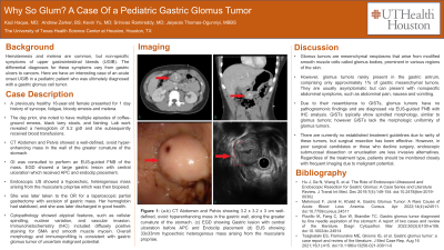
Case Description/Methods: A previously healthy 16-year-old female presented for 1 day history of syncope, fatigue, bloody emesis and melena. She noted to have multiple episodes of coffee-ground emesis, black tarry stools, and fainting prior to being admitted. She had a hemoglobin of 5.2 and received blood transfusions. CT Abdomen and Pelvis showed a well-defined, ovoid hyperenhancing mass in the wall of the greater curvature of the stomach. GI was consulted to perform an EUS-guided FNB of the mass. EGD showed a large gastric lesion with central ulceration status-post APC and endoclip placement. Endoscopic US showed a hypoechoic, heterogenous mass arising from the muscularis propriae which was biopsied. She was later taken for a laparoscopic partial gastrectomy with excision of gastric mass. She was discharged with resolution of symptoms. Cytopathology showed atypical features, such as cellular spindling, nuclear variation, and vascular invasion. Immunohistochemistry (IHC) included diffusely positive staining for SMA and smooth muscle myosin. Overall morphology and immunoprofiling is consistent with gastric glomus tumor of uncertain malignant potential.
Discussion: Glomus tumors are mesenchymal neoplasms that arise from modified smooth muscle cells called glomus bodies, prominent in various regions of the skin. However, glomus tumors rarely present in the gastric antrum, comprising only approximately 1% of gastric mesenchymal tumors. They are usually asymptomatic but can present with nonspecific abdominal symptoms. Due to their resemblance to GISTs, glomus tumors have no pathognomonic findings and are diagnosed via EUS-guided FNB with IHC analysis. GISTs typically show spindled morphology, similar to glomus tumors; however GISTs lack the morphologic uniformity of glomus tumors. There are currently no established treatment guidelines due to rarity of these tumors, but surgical resection has been effective. However, in poor surgical candidates or those who decline surgery, endoscopic submucosal dissection or enucleation are less invasive alternatives. Regardless of the treatment type, patients should be monitored closely with frequent imaging due to malignant potential.

Figure: Figure 1: (a-b) CT Abdomen and Pelvis showing 3.2 x 3.2 x 3 cm well-defined, ovoid hyperenhancing mass in the gastric wall, along the greater curvature of the stomach. (c) EGD showing Gastric lesion with central ulceration. (d) EUS showing 33x33mm hypoechoic heterogenous mass arising from the muscularis propriae.
Disclosures:
Kazi Haque indicated no relevant financial relationships.
Andrew Zarker indicated no relevant financial relationships.
Kevin Yu indicated no relevant financial relationships.
Jaiyeola Thomas-Ogunniyi indicated no relevant financial relationships.
Srinivas Ramireddy indicated no relevant financial relationships.
Kazi Haque, MD1, Andrew W. Zarker, BS1, Kevin Yu, MD2, Jaiyeola Thomas-Ogunniyi, MBBS1, Srinivas Ramireddy, MD1. P1189 - Why so Glum? A Case of a Pediatric Gastric Glomus Tumor, ACG 2023 Annual Scientific Meeting Abstracts. Vancouver, BC, Canada: American College of Gastroenterology.

