Sunday Poster Session
Category: Small Intestine
P1271 - Anemia: An Atypical Presentation of Gallstone Ileus
Sunday, October 22, 2023
3:30 PM - 7:00 PM PT
Location: Exhibit Hall

Has Audio

Shahina Saeed, DO
South Nassau Communities Hospital
Oceanside, NY
Presenting Author(s)
Shahina Saeed, DO1, Mahnoor Khan, DO2, Pranay Srivastava, MD3
1South Nassau Communities Hospital, Oceanside, NY; 2ISMMS Mt. Sinai South Nassau, Oceanside, NY; 3Mount Sinai South Nassau, Oceanside, NY
Introduction: Gallstone ileus is a rare complication of cholelithiasis, occurring in a fewer than 0.5% of patients who present with mechanical small bowel obstruction. Gallstone ileus typically occurs after an impaction of a gallstone in the ileum after being passed through a biliary-enteric fistula. The classic clinical presentation of gallstone ileus is an older woman with episodic subacute obstruction. Here we present a case of a patient who initially presented with symptoms of anemia, and she was incidentally noted to have a gallstone ileus.
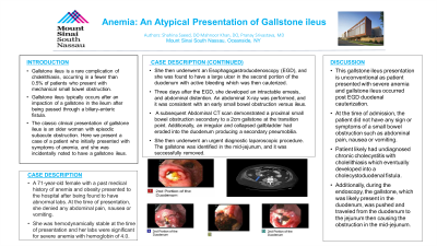
Case Description/Methods: A 71 year-old female with a past medical history of anemia and obesity presented to the hospital after being found to have abnormal labs. At the time of presentation, she denied any abdominal pain, nausea or vomiting. She was hemodynamically stable at the time of presentation and her labs were significant for severe anemia with hemoglobin of 4.0. She then underwent an Esophagogastroduodenoscopy (EGD), and she was found to have a large ulcer in the second portion of the duodenum with active bleeding which was then cauterized. Three days after the EGD, she developed an intractable emesis, and abdominal distention. An abdominal X-ray was performed, and it was consistent with an early small bowel obstruction versus ileus. A subsequent Abdominal CT scan demonstrated a proximal small bowel obstruction secondary to a 2cm gallstone at the transition point. Additionally, an irregular and collapsed gallbladder had eroded into the duodenum producing a secondary pneumobilia. She then under went an urgent diagnostic laparoscopic procedure. The gallstone was identified in the mid-jejunum, and it was successfully removed.
Discussion: This gallstone ileus presentation is unconventional as patient presented with severe anemia and gallstone ileus occurred post EGD duodenal cauterization. At the time of admission, the patient did not have any sign or symptoms of a small bowel obstruction such as abdominal pain, nausea or vomiting. Patient likely had undiagnosed chronic cholecystitis with cholelithiasis which eventually developed into a cholecystoduodenal fistula. Additionally, during the endoscopy, the gallstone, which was likely present in the duodenum, was pushed and traveled from the duodenum to the jejunum then causing the obstruction in the mid-jejunum.

Disclosures:
Shahina Saeed, DO1, Mahnoor Khan, DO2, Pranay Srivastava, MD3. P1271 - Anemia: An Atypical Presentation of Gallstone Ileus, ACG 2023 Annual Scientific Meeting Abstracts. Vancouver, BC, Canada: American College of Gastroenterology.
1South Nassau Communities Hospital, Oceanside, NY; 2ISMMS Mt. Sinai South Nassau, Oceanside, NY; 3Mount Sinai South Nassau, Oceanside, NY
Introduction: Gallstone ileus is a rare complication of cholelithiasis, occurring in a fewer than 0.5% of patients who present with mechanical small bowel obstruction. Gallstone ileus typically occurs after an impaction of a gallstone in the ileum after being passed through a biliary-enteric fistula. The classic clinical presentation of gallstone ileus is an older woman with episodic subacute obstruction. Here we present a case of a patient who initially presented with symptoms of anemia, and she was incidentally noted to have a gallstone ileus.
Case Description/Methods: A 71 year-old female with a past medical history of anemia and obesity presented to the hospital after being found to have abnormal labs. At the time of presentation, she denied any abdominal pain, nausea or vomiting. She was hemodynamically stable at the time of presentation and her labs were significant for severe anemia with hemoglobin of 4.0. She then underwent an Esophagogastroduodenoscopy (EGD), and she was found to have a large ulcer in the second portion of the duodenum with active bleeding which was then cauterized. Three days after the EGD, she developed an intractable emesis, and abdominal distention. An abdominal X-ray was performed, and it was consistent with an early small bowel obstruction versus ileus. A subsequent Abdominal CT scan demonstrated a proximal small bowel obstruction secondary to a 2cm gallstone at the transition point. Additionally, an irregular and collapsed gallbladder had eroded into the duodenum producing a secondary pneumobilia. She then under went an urgent diagnostic laparoscopic procedure. The gallstone was identified in the mid-jejunum, and it was successfully removed.
Discussion: This gallstone ileus presentation is unconventional as patient presented with severe anemia and gallstone ileus occurred post EGD duodenal cauterization. At the time of admission, the patient did not have any sign or symptoms of a small bowel obstruction such as abdominal pain, nausea or vomiting. Patient likely had undiagnosed chronic cholecystitis with cholelithiasis which eventually developed into a cholecystoduodenal fistula. Additionally, during the endoscopy, the gallstone, which was likely present in the duodenum, was pushed and traveled from the duodenum to the jejunum then causing the obstruction in the mid-jejunum.

Figure: Axial CT scan of the Abdomen with intravenous and gastrointestinal positive contrast. Showing multiple dilated Jejunal bowel loops. Abrupt caliber transition arises from obstructing gallstone
Disclosures:
Shahina Saeed indicated no relevant financial relationships.
Mahnoor Khan indicated no relevant financial relationships.
Pranay Srivastava indicated no relevant financial relationships.
Shahina Saeed, DO1, Mahnoor Khan, DO2, Pranay Srivastava, MD3. P1271 - Anemia: An Atypical Presentation of Gallstone Ileus, ACG 2023 Annual Scientific Meeting Abstracts. Vancouver, BC, Canada: American College of Gastroenterology.
